bsm-54036R [Primary Antibody]
Rabbit  Anti-Periostin Recombinant Rabbit mAb   Antibody
www.bioss.com.cn
sales@bioss.com.cn
techsupport@bioss.com.cn
400-901-9800
DATASHEET

Host: Rabbit

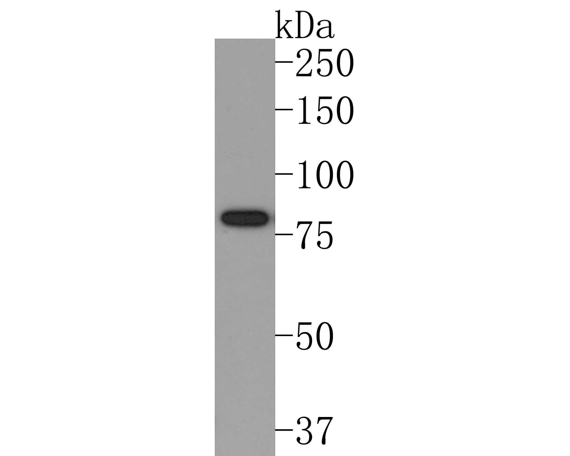
Target Protein: Periostin Recombinant Rabbit mAb

IR: Immunogen Range:15-54

Clonality:

Isotype: IgG

Entrez Gene: 10631

Swiss Prot: Q15063

Source: A synthesized peptide derived from human Periostin:15-54 

Purification: affinity purified by Protein A

Storage: 0.01M TBS (pH7.4) with 1% BSA, 0.02% Proclin300 and 50% Glycerol. Shipped at 4℃. Store at -20℃ for one year. Avoid repeated freeze/thaw cycles.

Background: Periostin is a disulfide linked 90 kDa, 811 amino acid protein originally isolated as a osteoblast-specific factor that functions as a cell adhesion molecule for preosteoblasts and is thought to be involved in osteoblast recruitment, attachment and spreading. Additionally, periostin expression has previously been shown to be significantly increased by both transforming growth factor beta 1(TGF beta 1) and bone morphogenetic protein (BMP2). Periostin has a typical signal sequence, followed by a cysteine-rich domain, a fourfold repeated domain and a C-terminal domain. The fourfold repeated domain of OSF2 shows homology with the insect protein fasciclin. Periostin mRNA is expressed in the developing mouse embryonic and fetal heart, and that it is localized to the endocardial cushions that ultimately divide the primitive heart tube into a four-chambered heart. Abnormal expression of periostin is also linked to angiogenesis and metastatsis in epithelial tumors.

Size: 50ul

Concentration: 1mg/ml

Applications: WB=1:500-2000,IHC-P=1:100-500,IHC-F=1:100-500,IF=1:100-500,Flow-Cyt=1:20-50

Cross Reactive Species: Human

For research use only. Not intended for diagnostic or therapeutic use.

VALIDATION IMAGES
Western blot analysis of Periostin on human brain tissue lysates. Proteins were transferred to a PVDF membrane and blocked with 5% NFDM/TBST in PBS for 1 hour at room temperature. The primary antibody (bsm-54036R, 1/500) was used in 5% NFDM/TBST at room temperature for 2 hours. Goat Anti-Rabbit IgG - HRP Secondary Antibody at 1:200,000 dilution was used for 1 hour at room temperature. Predicted band size: 93 kDa Observed band size: 80 kDa
Paraformaldehyde-fixed, paraffin embedded Human Esophagus Cancer; Antigen retrieval by boiling in sodium citrate buffer (pH6.0) for 15 min; Antibody incubation with Periostin Monoclonal Antibody, Unconjugated(bsm-54036R) at 1:200 overnight at 4°C, followed by conjugation to the bs-0295G-HRP and DAB (C-0010) staining.
Paraformaldehyde-fixed, paraffin embedded Human Stomach; Antigen retrieval by boiling in sodium citrate buffer (pH6.0) for 15 min; Antibody incubation with Periostin Monoclonal Antibody, Unconjugated(bsm-54036R) at 1:200 overnight at 4°C, followed by conjugation to the bs-0295G-HRP and DAB (C-0010) staining.
Paraformaldehyde-fixed, paraffin embedded Human Breast; Antigen retrieval by boiling in sodium citrate buffer (pH6.0) for 15 min; Antibody incubation with Periostin Monoclonal Antibody, Unconjugated(bsm-54036R) at 1:200 overnight at 4°C, followed by conjugation to the bs-0295G-HRP and DAB (C-0010) staining.
Paraformaldehyde-fixed, paraffin embedded Human Colon; Antigen retrieval by boiling in sodium citrate buffer (pH6.0) for 15 min; Antibody incubation with Periostin Monoclonal Antibody, Unconjugated(bsm-54036R) at 1:200 overnight at 4°C, followed by conjugation to the bs-0295G-HRP and DAB (C-0010) staining.
Paraformaldehyde-fixed, paraffin embedded Human Lung Cancer; Antigen retrieval by boiling in sodium citrate buffer (pH6.0) for 15 min; Antibody incubation with Periostin Monoclonal Antibody, Unconjugated(bsm-54036R) at 1:200 overnight at 4°C, followed by conjugation to the bs-0295G-HRP and DAB (C-0010) staining.
Flow cytometric analysis of Periostin was done on 293T cells. The cells were fixed, permeabilized and stained with the primary antibody (bsm-54036R, 1/50) (blue). After incubation of the primary antibody at room temperature for an hour, the cells were stained with a Alexa Fluor®488 conjugate-Goat anti-Rabbit IgG Secondary antibody at 1/1,000 dilution for 30 minutes.Unlabelled sample was used as a control (cells without incubation with primary antibody; red).
 
PRODUCT SPECIFIC PUBLICATIONS