sales@bioss.com.cn
techsupport@bioss.com.cn
400-901-9800
Host: Rabbit
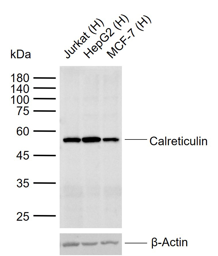
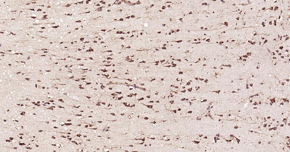
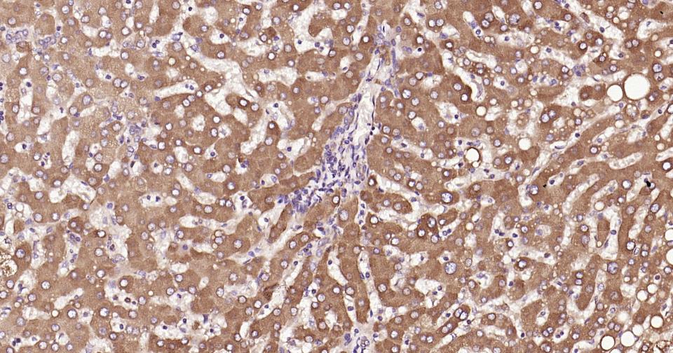
Target Protein: Calreticulin Rabbit pAb
IR: Immunogen Range:101-200/417
Clonality: Polyclonal
Isotype: IgG
Entrez Gene: 811
Swiss Prot: P27797
Source: KLH conjugated synthetic peptide derived from human Calreticulin:101-200/417
Purification: affinity purified by Protein A
Storage: 0.01M TBS (pH7.4) with 1% BSA, 0.02% Proclin300 and 50% Glycerol. Shipped at 4℃. Store at -20℃ for one year. Avoid repeated freeze/thaw cycles.
Background: Calreticulin is a highly conserved chaperone protein which resides primarily in the endoplasmic reticulum, and is involved in a variety of cellular processes, among them, cell adhesion. Additionally, it functions in protein folding quality control and calcium homeostasis. Calreticulin is also found in the nucleus, suggesting that it may have a role in transcription regulation. Systemic lupus erythematosus is associated with increased autoantibody titers against calreticulin. Recurrent mutations in calreticulin have been linked to various neoplasms, including the myeloproliferative type.[provided by RefSeq, May 2020]
Size: 50ul
Concentration: 1mg/ml
Applications: WB=1:500-2000,IHC-P=1:100-500,IHC-F=1:100-500,IF=1:100-500,ELISA=1:5000-10000
Cross Reactive Species: Human
For research use only. Not intended for diagnostic or therapeutic use.