bs-1534R [Primary Antibody]
Rabbit  Anti-MAP1LC3A  Polyclonal Antibody
www.bioss.com.cn
sales@bioss.com.cn
techsupport@bioss.com.cn
400-901-9800
DATASHEET

Host: Rabbit

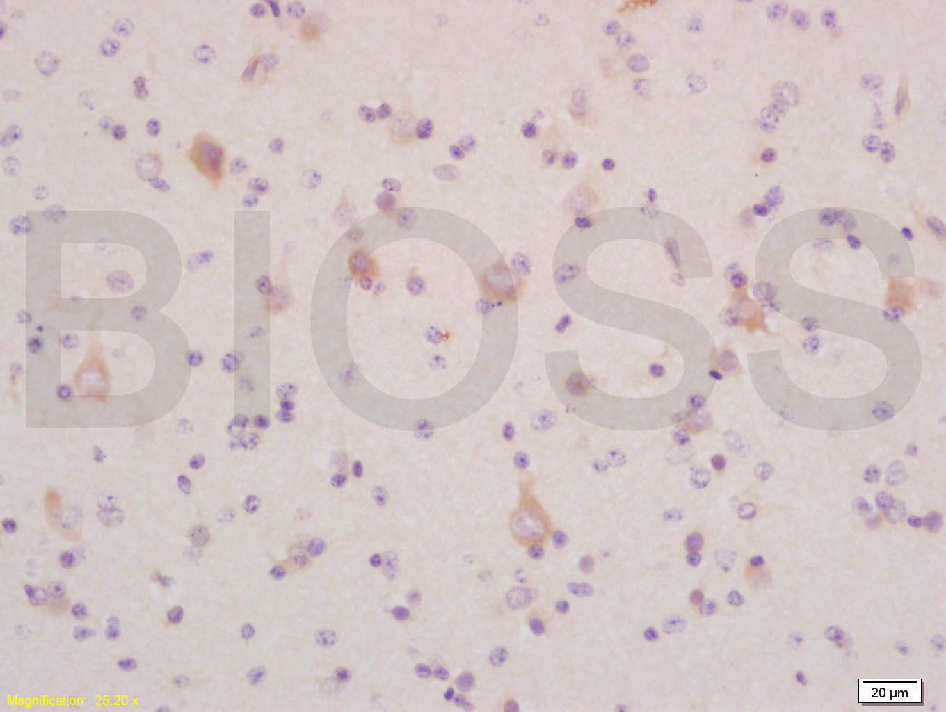
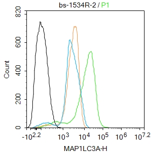
Target Protein: MAP1LC3A

IR: Immunogen Range:75-121/121

Clonality: Polyclonal

Isotype: IgG

Entrez Gene: 84557

Swiss Prot: Q9H492

Source: KLH conjugated synthetic peptide derived from human MAP1LC3A:75-121/121 

Purification: affinity purified by Protein A

Storage: 0.01M TBS (pH7.4) with 1% BSA, 0.02% Proclin300 and 50% Glycerol. Shipped at 4℃. Store at -20℃ for one year. Avoid repeated freeze/thaw cycles.

Background: Microtubule-associated MAPILC3A constitutes nearly half of the mass of all the microtubule associated proteins that copurify with brain microtubules. MAP1LC3A is one of three human orthologs of the rat Map1LC3, (named MAP1LC3A, MAP1LC3B, and MAP1LC3C). The three isoforms of human MAP1LC3 exhibit distinct expression patterns in different human tissues and also differ in their post-translation modifications. MAP1LC3A and MAP1LC3C are produced by the proteolytic cleavage after the conserved C-terminal Gly residue; MAP1LC3B does not undergo C-terminal cleavage and exists in a single modified form.

Size: 50ul

Concentration: 1mg/ml

Applications: IHC-P=1:100-500,IHC-F=1:100-500,Flow-Cyt=2μg/Test,IF=1:100-500,ELISA=1:5000-10000

Cross Reactive Species: Human,Mouse (predicted: Rat,Pig,Cow,Chicken)

For research use only. Not intended for diagnostic or therapeutic use.

VALIDATION IMAGES
Tissue/cell: human glioma tissue; 4% Paraformaldehyde-fixed and paraffin-embedded; Antigen retrieval: citrate buffer ( 0.01M, pH 6.0 ), Boiling bathing for 15min; Block endogenous peroxidase by 3% Hydrogen peroxide for 30min; Blocking buffer (normal goat serum,C-0005) at 37℃ for 20 min; Incubation: Anti-LC3 α/MAP1A/MAP LC3 Alpha/Beta Polyclonal Antibody, Unconjugated (bs-1534R) 1:200, overnight at 4°C, followed by conjugation to the secondary antibody(SP-0023) and DAB(C-0010) staining
Blank control (blue line): Hela (blue). Primary Antibody (green line): Rabbit Anti-MAP1LC3A antibody (bs-1534R) Dilution: 0.2μg /10^6 cells; Isotype Control Antibody (orange line): Rabbit IgG . Secondary Antibody (white blue line): Goat anti-rabbit IgG-PE Dilution: 1μg /test. Protocol The cells were fixed with 70% ethanol (Overmight at 4℃) and then permeabilized with 90% ice-cold methanol for 30 min at -20℃.Cells stained with Primary Antibody for 30 min at room temperature. The cells were then incubated in 1 X PBS/2%BSA/10% goat serum to block non-specific protein-protein interactions followed by the antibody for 15 min at room temperature. The secondary antibody used for 40 min at room temperature. Acquisition of 20,000 events was performed.
Blank control: RAW264.7. Primary Antibody (green line): Rabbit Anti-MAP1LC3A antibody (bs-1534R) Dilution: 2ug/Test; Secondary Antibody : Goat anti-rabbit IgG-FITC Dilution: 0.5ug/Test. Protocol The cells were fixed with 4% PFA (10min at room temperature)and then permeabilized with 0.1% PBST for 20 min at room temperature.The cells were then incubated in 5%BSA to block non-specific protein-protein interactions for 30 min at room temperature .Cells stained with Primary Antibody for 30 min at room temperature. The secondary antibody used for 40 min at room temperature. Acquisition of 20,000 events was performed.
 
PRODUCT SPECIFIC PUBLICATIONS