bs-1238R [Primary Antibody]
Rabbit  Anti-HO-2 Polyclonal Antibody
www.bioss.com.cn
sales@bioss.com.cn
techsupport@bioss.com.cn
400-901-9800
DATASHEET

Host: Rabbit

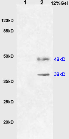
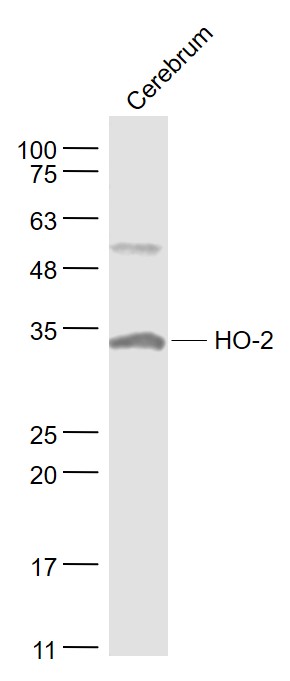
Target Protein: HO-2

IR: Immunogen Range:31-150/316

Clonality: Polyclonal

Isotype: IgG

Entrez Gene: 3163

Swiss Prot: P30519

Source: KLH conjugated synthetic peptide derived from human HO-2:31-150/316 

Purification: affinity purified by Protein A

Storage: 0.01M TBS(pH7.4) with 1% BSA, 0.03% Proclin300 and 50% Glycerol. Shipped at 4℃. Store at -20 °C for one year. Avoid repeated freeze/thaw cycles.

Background: Heme oxygenase cleaves the heme ring at the alpha methene bridge to form biliverdin. Biliverdin is subsequently converted to bilirubin by biliverdin reductase. Under physiological conditions, the activity of heme oxygenase is highest in the spleen, where senescent erythrocytes are sequestrated and destroyed. Heme oxygenase 2 could be implicated in the production of carbon monoxide in brain where it could act as a neurotransmitter. Summary: catalyzes the conversion of heme to biliverdin; involved in cellular response to oxidative stress [SUBCELLULAR LOCATION] Microsome. Endoplasmic reticulum. [INDUCTION] Heme oxygenase 2 activity is non-inducible. [SIMILARITY] Belongs to the heme oxygenase family.

Size: 50ul

Concentration: 1mg/ml

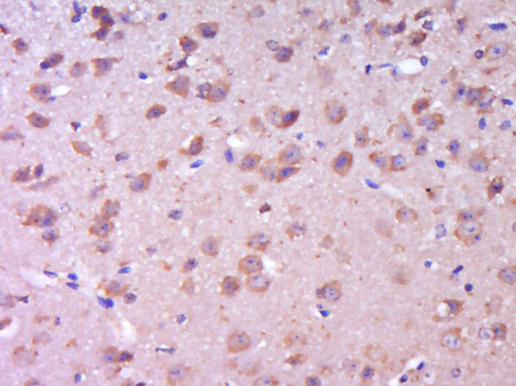
Applications: WB=1:500-2000,IHC-P=1:100-500,IHC-F=1:100-500,Flow-Cyt=0.2ug/test,IF=1:100-500,ELISA=1:5000-10000

Cross Reactive Species: Human,Mouse,Rat

For research use only. Not intended for diagnostic or therapeutic use.

VALIDATION IMAGES
Sample: Brain(Rat)lysate at 30ug; Liver(Rat) lysate at 30ug; Primary: Anti-HO-2 (bs-1238R) at 1:200 dilution; Secondary: HRP conjugated Goat Anti-Rabbit IgG(bs-0295G-HRP) at 1: 3000 dilution; Predicted band size : 38kD Observed band size : 38kD, 48kD
Sample: Cerebrum (Mouse) Lysate at 40 ug Primary: Anti- HO-2 (bs-1238R) at 1/1000 dilution Secondary: IRDye800CW Goat Anti-Rabbit IgG at 1/20000 dilution Predicted band size: 36 kD Observed band size: 33 kD
Paraformaldehyde-fixed, paraffin embedded (Mouse brain); Antigen retrieval by boiling in sodium citrate buffer (pH6.0) for 15min; Block endogenous peroxidase by 3% hydrogen peroxide for 20 minutes; Blocking buffer (normal goat serum) at 37°C for 30min; Antibody incubation with (HO-2) Polyclonal Antibody, Unconjugated (bs-1238R) at 1:400 overnight at 4°C, followed by operating according to SP Kit(Rabbit) (sp-0023) instructionsand DAB staining.
U-937 cells were fixed with 4% PFA for 10min at room temperature,permeabilized with 20% PBST for 20 min at room temperature, and incubated in 5% BSA blocking buffer for 30 min at room temperature. Cells were then stained with HO-2 Antibody(bs-1238R)at 1:500 dilution in blocking buffer and incubated for 30 min at room temperature, washed twice with 2%BSA in PBS, followed by secondary antibody incubation for 40 min at room temperature. Acquisitions of 20,000 events were performed. Cells stained with primary antibody (green), and isotype control (orange).
 
PRODUCT SPECIFIC PUBLICATIONS