bsm-54023R [Primary Antibody]
Rabbit  Anti-LXR alpha   Antibody
www.bioss.com.cn
sales@bioss.com.cn
techsupport@bioss.com.cn
400-901-9800
DATASHEET

Host: Rabbit

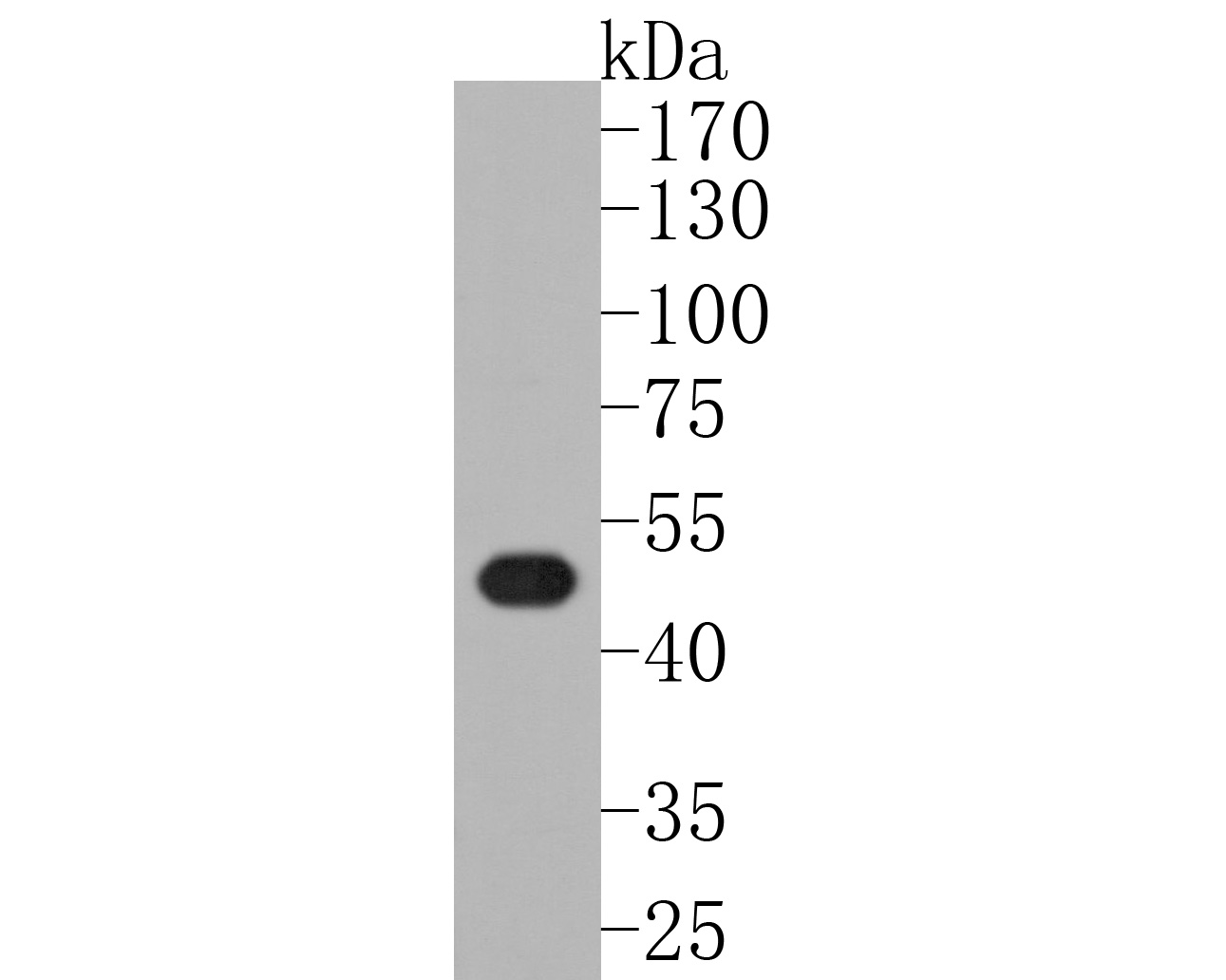
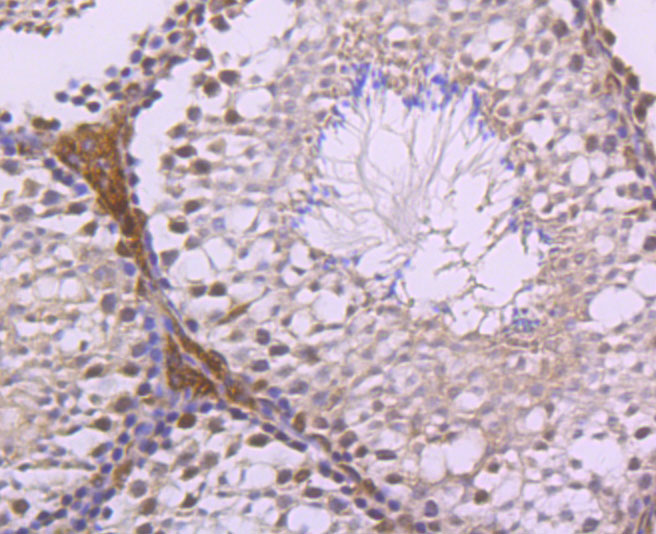
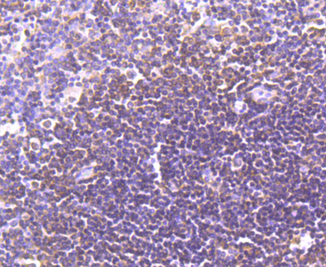
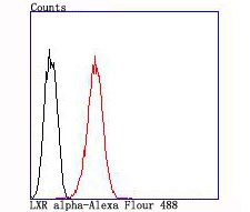
Target Protein: LXR alpha

IR: Immunogen Range:60-110/447

Clonality:

Isotype:

Entrez Gene: 10062

Swiss Prot: Q13133

Source: KLH conjugated synthetic peptide derived from human LXR alpha:60-110/447 

Purification: affinity purified by Protein A

Storage: 0.01M TBS (pH7.4) with 1% BSA, 0.02% Proclin300 and 50% Glycerol. Shipped at 4℃. Store at -20℃ for one year. Avoid repeated freeze/thaw cycles.

Background: Peroxisome proliferators include hypolipidemic drugs, herbicides, leukotriene antagonists, and plasticizers; this term arises because they induce an increase in the size and number of peroxisomes. Peroxisomes are subcellular organelles found in plants and animals that contain enzymes for respiration and for cholesterol and lipid metabolism. The action of peroxisome proliferators is thought to be mediated via specific receptors, called PPARs, which belong to the steroid hormone receptor superfamily. PPARs affect the expression of target genes involved in cell proliferation, cell differentiation and in immune and inflammation responses. Three closely related subtypes (alpha, beta/delta, and gamma) have been identified. This gene encodes the subtype PPAR-alpha, which is a nuclear transcription factor. Multiple alternatively spliced transcript variants have been described for this gene, although the full-length nature of only two has been determined. [provided by RefSeq, Jul 2008].

Size: 50ul

Concentration: 1mg/ml

Applications: WB=1:500-1000,IHC-P=1:50-200,IHC-F=1:400-800,Flow-Cyt=1:50,ICC/IF=1:50,IF=1:100-500

Cross Reactive Species: Human,Mouse (predicted: Rat)

For research use only. Not intended for diagnostic or therapeutic use.

VALIDATION IMAGES
Sample: Lane 1: mouse colon tissue lysates Primary: Anti-LXR alpha (bsm-54023R) at 1:500 dilution Secondary: Goat Anti-Rabbit IgG - HRP at 1:5000 dilution Predicted band size: 50 kD Observed band size: 50 kD
Sample: Lane 1: human liver tissue lysates Primary: Anti-LXR alpha (bsm-54023R) at 1:500 dilution Secondary: Goat Anti-Rabbit IgG - HRP at 1:5000 dilution Predicted band size: 50 kD Observed band size: 53 kD
Paraformaldehyde-fixed, paraffin embedded (mouse testis tissue); Antigen retrieval by boiling in sodium citrate buffer (pH6.0) for 15min; Block endogenous peroxidase by 3% hydrogen peroxide for 20 minutes; Blocking buffer (normal goat serum) at 37°C for 30min; Antibody incubation with (LXR alpha) Monoclonal Antibody, Unconjugated (bsm-54023R) at 1:50 overnight at 4°C, followed by operating according to SP Kit(Rabbit) (sp-0023) instructionsand DAB staining.
Paraformaldehyde-fixed, paraffin embedded (human spleen tissue); Antigen retrieval by boiling in sodium citrate buffer (pH6.0) for 15min; Block endogenous peroxidase by 3% hydrogen peroxide for 20 minutes; Blocking buffer (normal goat serum) at 37°C for 30min; Antibody incubation with (LXR alpha) Monoclonal Antibody, Unconjugated (bsm-54023R) at 1:50 overnight at 4°C, followed by operating according to SP Kit(Rabbit) (sp-0023) instructionsand DAB staining.
Paraformaldehyde-fixed, paraffin embedded (human placenta tissue); Antigen retrieval by boiling in sodium citrate buffer (pH6.0) for 15min; Block endogenous peroxidase by 3% hydrogen peroxide for 20 minutes; Blocking buffer (normal goat serum) at 37°C for 30min; Antibody incubation with (LXR alpha) Monoclonal Antibody, Unconjugated (bsm-54023R) at 1:50 overnight at 4°C, followed by operating according to SP Kit(Rabbit) (sp-0023) instructionsand DAB staining.
HepG2 cell; 4% Paraformaldehyde-fixed; Triton X-100 at room temperature for 20 min; Blocking buffer (normal goat serum, C-0005) at 37°C for 20 min; Antibody incubation with (LXR alpha) monoclonal Antibody, Unconjugated (bsm-54023R) 1:50, 90 minutes at 37°C; followed by a conjugated Goat Anti-Rabbit IgG antibody at 37°C for 90 minutes, DAPI (blue, C02-04002) was used to stain the cell nuclei.
Blank control:HepG2 . Primary Antibody (green line): Rabbit Anti-LXR alpha antibody (bsm-54023R) Dilution: 1:50; Isotype Control Antibody (orange line): Rabbit IgG . Secondary Antibody : Goat anti-rabbit IgG-AF488 Dilution: 1:1000. Protocol The cells were fixed with 4% PFA (10min at room temperature)and then permeabilized with 90% ice-cold methanol for 20 min at -20℃. The cells were then incubated in 5%BSA to block non-specific protein-protein interactions for 30 min at room temperature .Cells stained with Primary Antibody for 30 min at room temperature. The secondary antibody used for 40 min at room temperature. Acquisition of 20,000 events was performed.
 
PRODUCT SPECIFIC PUBLICATIONS