bs-55093R [Primary Antibody]
Rabbit  Anti-HDAC1  Polyclonal Antibody
www.bioss.com.cn
sales@bioss.com.cn
techsupport@bioss.com.cn
400-901-9800
DATASHEET

Host: Rabbit

Target Protein: HDAC1

IR: Immunogen Range:393-482/482

Clonality: Polyclonal

Isotype: IgG

Entrez Gene: 3065

Swiss Prot: Q13547

Source: Recombinant human HDAC1 :393-482/482 

Purification: affinity purified by Protein A

Storage: 0.01M TBS(pH7.4) with 1% BSA, 0.03% Proclin300 and 50% Glycerol. Store at -20 °C for one year. Avoid repeated freeze/thaw cycles. The lyophilized antibody is stable at room temperature for at least one month and for greater than a year when kept at -20°C. When reconstituted in sterile pH 7.4 0.01M PBS or diluent of antibody the antibody is stable for at least two weeks at 2-4 °C.

Background: Histone acetylation and deacetylation, catalyzed by multisubunit complexes, play a key role in the regulation of eukaryotic gene expression. The protein encoded by this gene belongs to the histone deacetylase/acuc/apha family and is a component of the histone deacetylase complex. It also interacts with retinoblastoma tumor-suppressor protein and this complex is a key element in the control of cell proliferation and differentiation. Together with metastasis-associated protein-2, it deacetylates p53 and modulates its effect on cell growth and apoptosis. [provided by RefSeq, Jul 2008]

Size: 50ul

Concentration: 1mg/ml

Applications: WB=1:500-2000,IHC-P=1:100-200,IHC-F=1:100-200,ICC/IF=1:100,IF=1:50-100

Cross Reactive Species: Human,Rat (predicted: Mouse)

For research use only. Not intended for diagnostic or therapeutic use.
VALIDATION IMAGES
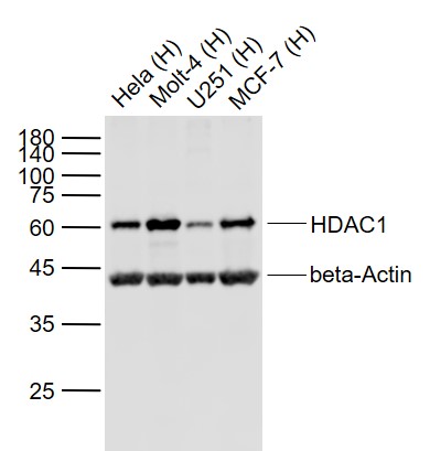
Sample: Lane 1: Hela (Human) Cell Lysate at 30 ug Lane 2: Molt-4 (Human) Cell Lysate at 30 ug Lane 3: U251 (Human) Cell Lysate at 30 ug Lane 4: MCF-7 (Human) Cell Lysate at 30 ug Primary: Anti- HDAC1 (bs-55093R) at 1/1000 dilution Anti- beta-Actin (bs-0061R) at 1/2000 dilution Secondary: IRDye800CW Goat Anti-Rabbit IgG at 1/20000 dilution Predicted band size: 62 kD Observed band size: 60 kD
Sample: Lane 1: 293T (Human) Cell Lysate at 25 ug Lane 2: HDAC1 knockout (KO) 293T (Human) Cell Lysate at 25 ug Primary: Anti-HDAC1 (bs-55093R) at 1/1000 dilution Secondary: HRP Goat Anti-Rabbit IgG (H+L) at 1:10000 dilution Predicted band size: 62 kD Observed band size: 65 kD
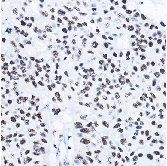
Paraformaldehyde-fixed, paraffin embedded (human colon); Antigen retrieval by boiling in sodium citrate buffer (pH6.0) for 15min; Block endogenous peroxidase by 3% hydrogen peroxide for 20 minutes; Blocking buffer (normal goat serum) at 37°C for 30min; Antibody incubation with (HDAC1) Polyclonal Antibody, Unconjugated (bs-55093R) at 1:50 overnight at 4°C, followed by operating according to SP Kit(Rabbit) (sp-0023) instructionsand DAB staining.
Paraformaldehyde-fixed, paraffin embedded (human lung cancer); Antigen retrieval by boiling in sodium citrate buffer (pH6.0) for 15min; Block endogenous peroxidase by 3% hydrogen peroxide for 20 minutes; Blocking buffer (normal goat serum) at 37°C for 30min; Antibody incubation with (HDAC1) Polyclonal Antibody, Unconjugated (bs-55093R) at 1:50 overnight at 4°C, followed by operating according to SP Kit(Rabbit) (sp-0023) instructionsand DAB staining.
Paraformaldehyde-fixed, paraffin embedded (rat spleen); Antigen retrieval by boiling in sodium citrate buffer (pH6.0) for 15min; Block endogenous peroxidase by 3% hydrogen peroxide for 20 minutes; Blocking buffer (normal goat serum) at 37°C for 30min; Antibody incubation with (HDAC1) Polyclonal Antibody, Unconjugated (bs-55093R) at 1:50 overnight at 4°C, followed by operating according to SP Kit(Rabbit) (sp-0023) instructionsand DAB staining.
L929 cell; 4% Paraformaldehyde-fixed; Triton X-100 at room temperature for 20 min; Blocking buffer (normal goat serum, C-0005) at 37°C for 20 min; Antibody incubation with (KO Validated)HDAC1 polyclonal Antibody, Unconjugated (bs-55093R) 1:200, 90 minutes at 37°C; followed by a conjugated Goat Anti-Rabbit IgG antibody at 37°C for 90 minutes, DAPI (blue, C02-04002) was used to stain the cell nuclei.
A549 cell; 4% Paraformaldehyde-fixed; Triton X-100 at room temperature for 20 min; Blocking buffer (normal goat serum, C-0005) at 37°C for 20 min; Antibody incubation with (KO Validated)HDAC1 polyclonal Antibody, Unconjugated (bs-55093R) 1:100, 90 minutes at 37°C; followed by a conjugated Goat Anti-Rabbit IgG antibody at 37°C for 90 minutes, DAPI (blue, C02-04002) was used to stain the cell nuclei.
 
PRODUCT SPECIFIC PUBLICATIONS