VALIDATION IMAGES
Sample:
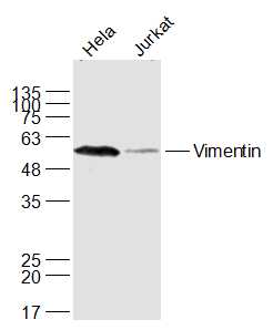
A549(Human) Cell Lysate at 30 ug
Jurkat(Human) Cell Lysate at 30 ug
Primary: Anti-Vimentin (bs-8533R) at 1/1000 dilution
Secondary: IRDye800CW Goat Anti-Rabbit IgG at 1/20000 dilution
Predicted band size: 51 kD
Observed band size: 53 kD
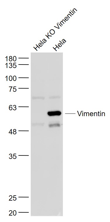
Sample:
Hela KO Vimentin (Human) Cell Lysate at 30 ug
Hela(Human) Cell Lysate at 30 ug
Primary: Anti- Vimentin (bs-8533R) at 1/1000 dilution
Secondary: IRDye800CW Goat Anti-Rabbit IgG at 1/20000 dilution
Predicted band size: 51 kD
Observed band size: 57 kD
Protein: lung(rabbit) lysate at 40ug;
Primary: rabbit Anti-Vimentin (bs-8533R) at 1:300;
Secondary: HRP conjugated Goat-Anti-rabbit IgG(bs-0295G-HRP) at 1: 5000;
Predicted band size: 51 kD
Observed band size: 51 kD
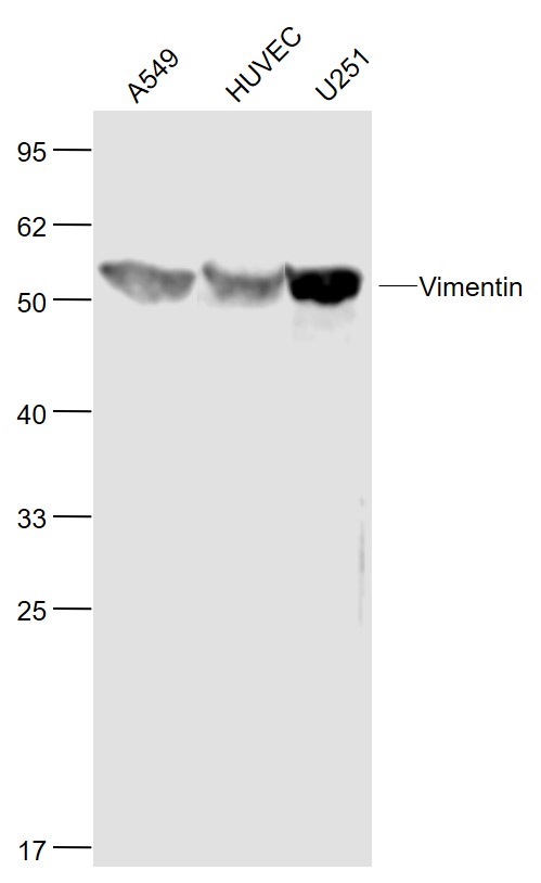
Sample:
A549(Human) Cell Lysate at 30 ug
HUVEC(Human) Cell Lysate at 30 ug
U251(Human) Cell Lysate at 30 ug
Primary: Anti-Vimentin (bs-8533R) at 1/1000 dilution
Secondary: IRDye800CW Goat Anti-Rabbit IgG at 1/20000 dilution
Predicted band size: 53 kD
Observed band size: 53 kD
Sample:
U-87MG (Human) Cell Lysate at 30 ug
NIH/3T3 (Mouse) Cell Lysate at 30 ug
Primary: Anti-Vimentin (bs-8533R) at 1/1000 dilution
Secondary: IRDye800CW Goat Anti-Rabbit IgG at 1/20000 dilution
Predicted band size: 51 kD
Observed band size: 53 kD
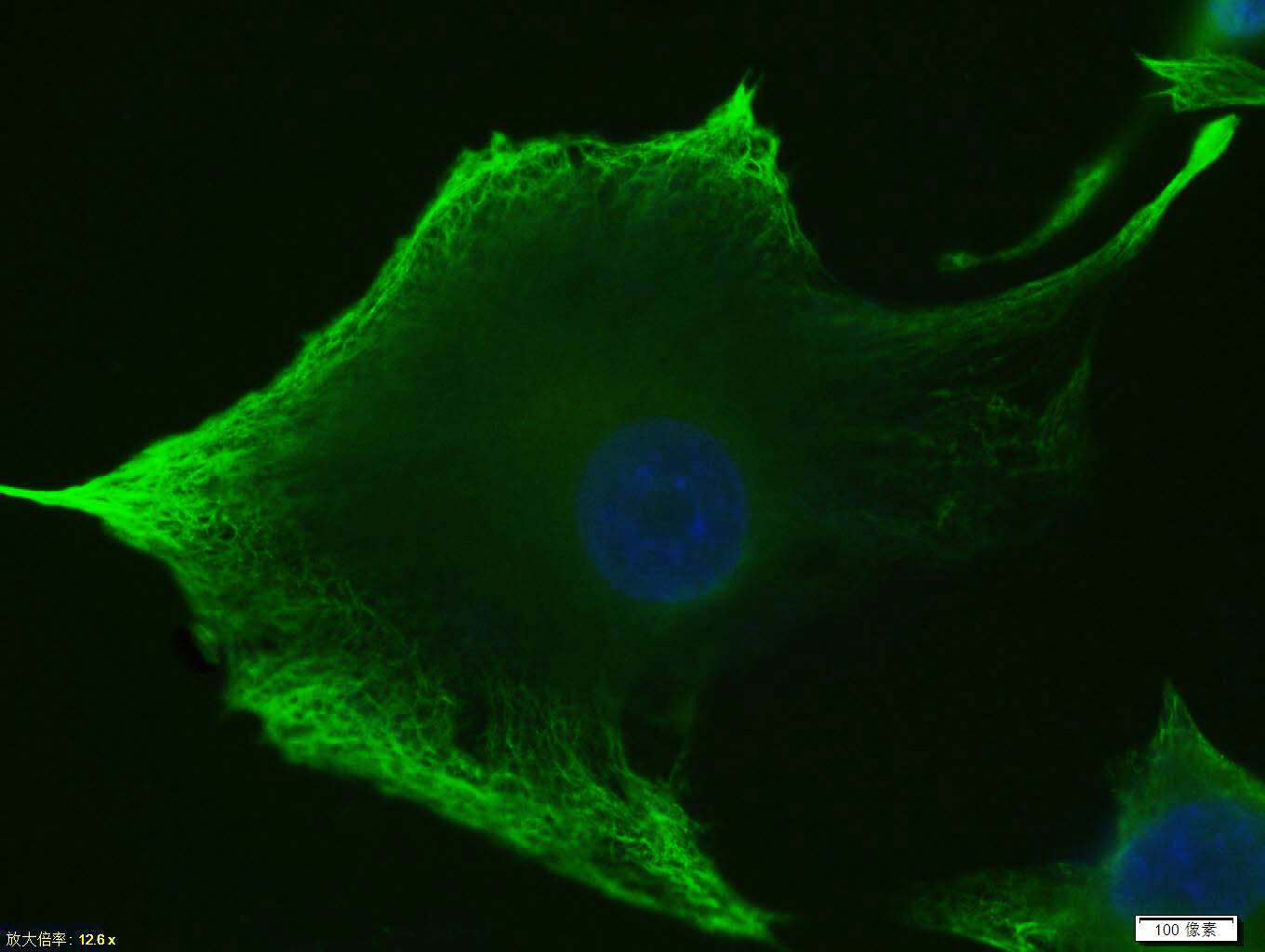
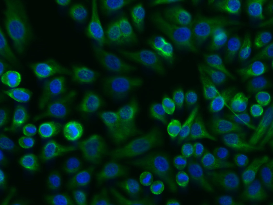
Tissue/cell: U-87MG cell; 4% Paraformaldehyde-fixed; Triton X-100 at room temperature for 20 min; Blocking buffer (normal goat serum, C-0005) at 37°C for 20 min; Antibody incubation with (Vimentin) Polyclonal Antibody, Unconjugated (bs-8533R)antibody (bs-0295G-FITC) at 37°C for 90 minutes, DAPI (blue, C02-04002) was used to stain the cell nuclei.
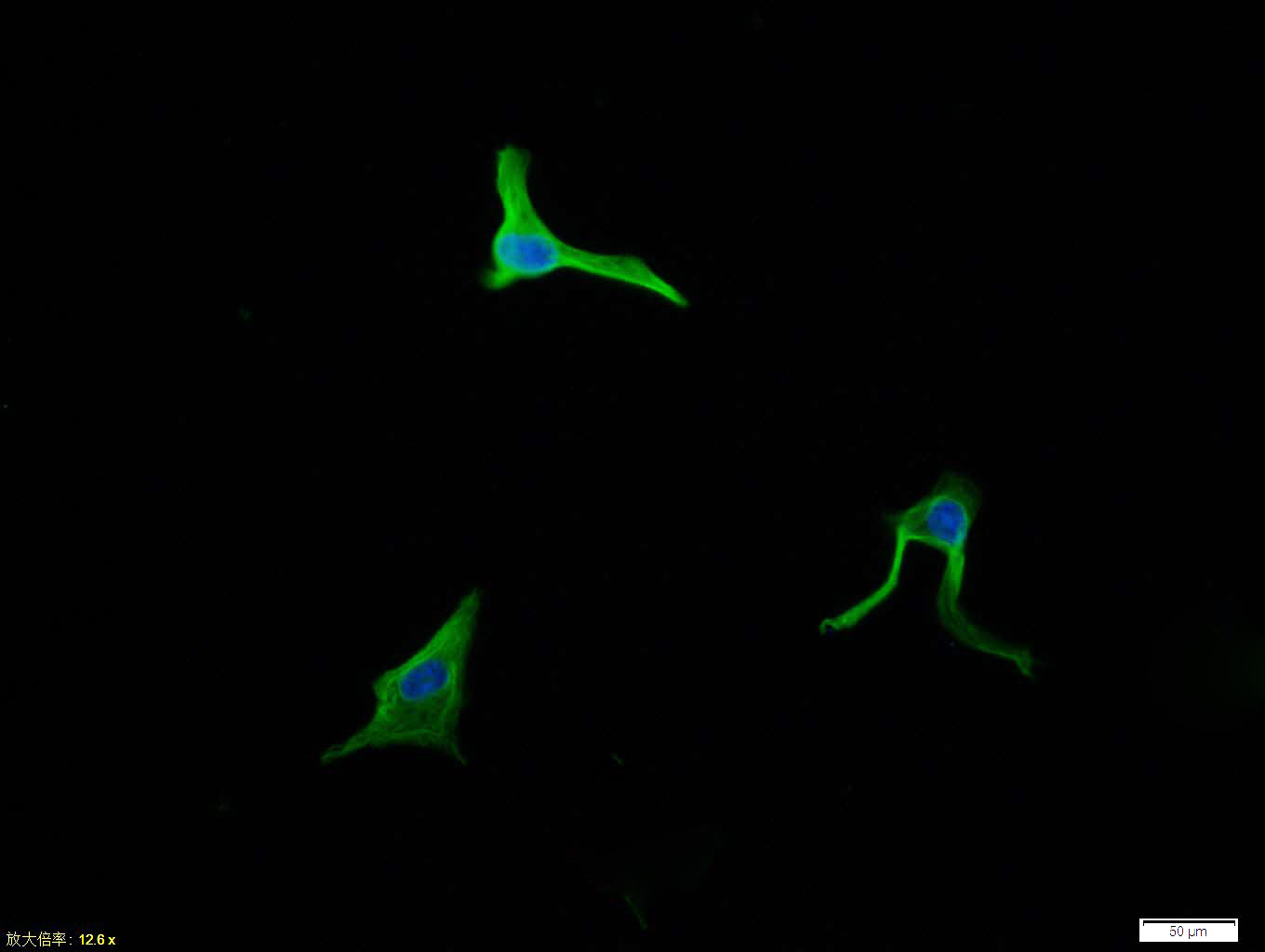
Tissue/cell: endothelial cells of umbilical artery;4% Paraformaldehyde-fixed;
Blocking buffer (normal goat serum,C-0005) at 37℃ for 20 min;
Incubation: Anti-Vimentin Polyclonal Antibody, Alexa Fluor 488 conjugated(bs-8533R-A488) 1:100, 60 minutes at 37℃. DAPI(5ug/ml,blue,C-0033) was used to stain the cell nuclei
Tissue/cell: U-87MG cell; 4% Paraformaldehyde-fixed; Triton X-100 at room temperature for 20 min; Blocking buffer (normal goat serum, C-0005) at 37°C for 20 min; Antibody incubation with (Vimentin) polyclonal Antibody, Unconjugated (bs-8533R) 1:100, 90 minutes at 37°C; followed by a conjugated Goat Anti-Rabbit IgG antibody at 37°C for 90 minutes, DAPI (blue, C02-04002) was used to stain the cell nuclei.
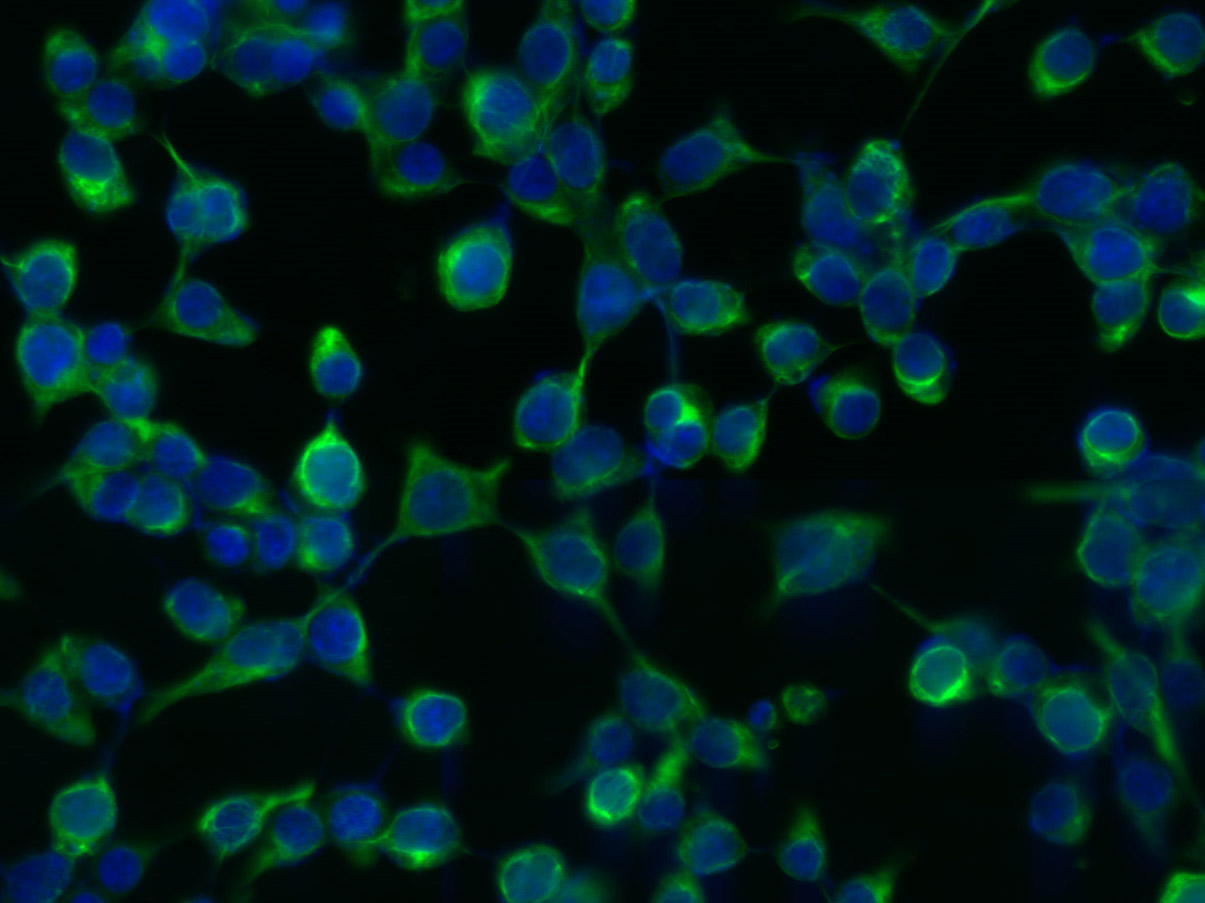
Tissue/cell: 293T cell; 4% Paraformaldehyde-fixed; Triton X-100 at room temperature for 20 min; Blocking buffer (normal goat serum, C-0005) at 37°C for 20 min; Antibody incubation with (Vimentin) Polyclonal Antibody, Unconjugated (bs-8533R) 1:200, 2 hours at 37°C; followed by a conjugated Goat Anti-Rabbit IgG antibody (bs-0295G-FITC) at 37°C for 90 minutes, DAPI (5ug/ml, blue, C-0033) was used to stain the cell nuclei.
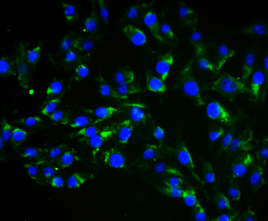
Tissue/cell: FHC cell; 4% Paraformaldehyde-fixed; Triton X-100 at room temperature for 20 min; Blocking buffer (normal goat serum, C-0005) at 37°C for 20 min; Antibody incubation with (Vimentin) Polyclonal Antibody, Unconjugated (bs-8533R) 1:200, 2 hours at 37°C; followed by a conjugated Goat Anti-Rabbit IgG antibody (bs-0295G-FITC) at 37°C for 90 minutes, DAPI (5ug/ml, blue, C-0033) was used to stain the cell nuclei.
Blank control:A549.
Primary Antibody (green line): Rabbit Anti-Vimentin antibody (bs-8533R)
Dilution: 1μg /10^6 cells;
Isotype Control Antibody (orange line): Rabbit IgG .
Secondary Antibody : Goat anti-rabbit IgG-AF488
Dilution: 1μg /test.
Protocol
The cells were fixed with 4% PFA (10min at room temperature)and then permeabilized with 90% ice-cold methanol for 20 min at -20℃. The cells were then incubated in 5%BSA to block non-specific protein-protein interactions for 30 min at room temperature .Cells stained with Primary Antibody for 30 min at room temperature. The secondary antibody used for 40 min at room temperature. Acquisition of 20,000 events was performed.