sales@bioss.com.cn
techsupport@bioss.com.cn
400-901-9800
Host: Rabbit
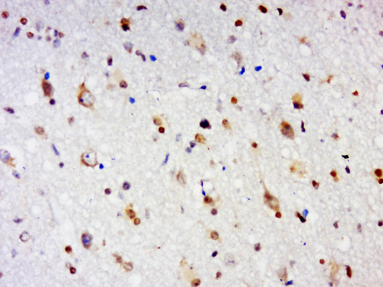
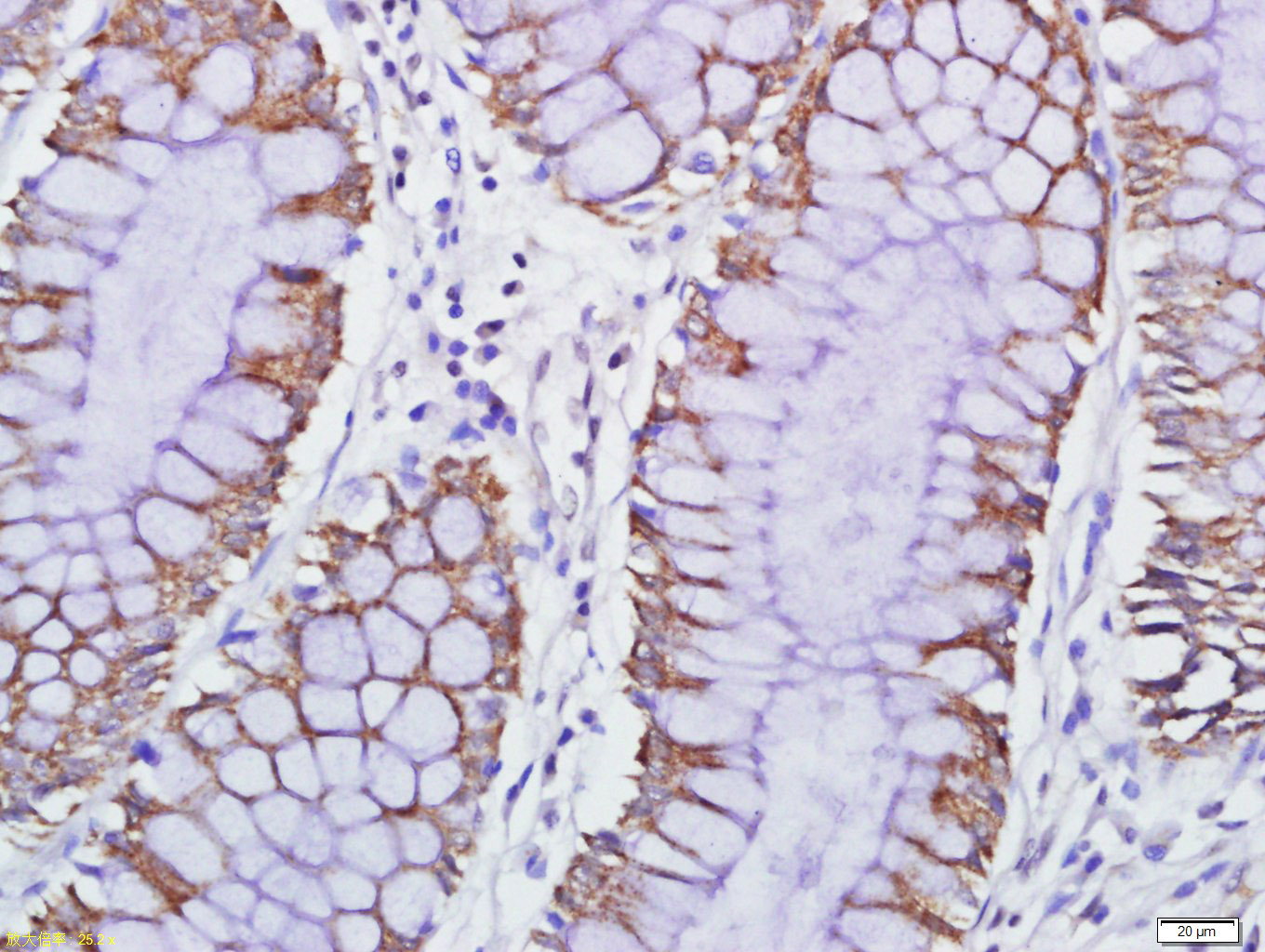
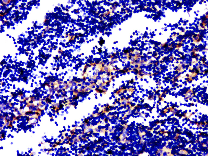
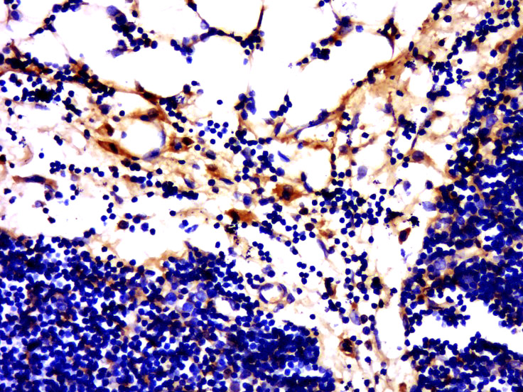
Target Protein: GPR84 Rabbit pAb
IR: Immunogen Range:21-120/369
Clonality: Polyclonal
Isotype: IgG
Entrez Gene: 53831
Swiss Prot: Q9NQS5
Source:
KLH conjugated synthetic peptide derived from human GPCR EX33 protein:21-120/369
Purification: affinity purified by Protein A
Storage: 0.01M TBS (pH7.4) with 1% BSA, 0.02% Proclin300 and 50% Glycerol. Shipped at 4℃. Store at -20℃ for one year. Avoid repeated freeze/thaw cycles.
Background: G protein-coupled receptors (GPCRs), also designated seven transmembrane (7TM) receptors and heptahelical receptors, are a protein family which interact with G proteins (heterotrimeric GTPases) to synthesize intracellular second messengers such as diacylglycerol, cyclic AMP, inositol phosphates, and calcium ions. Their diverse biological functions range from vision and olfaction to neuronal and endocrine signaling and are involved in many pathological conditions. G protein receptor 84 (GPR84), a member of the GCPR 1 family, is an orphan GCPR expressed in bone marrow, brain, heart, muscle, colon, thymus, spleen, kidney, liver, placenta, intestine, lung and peripheral blood leukocytes. In activated T cells, GPR84 regulates early interleukin-4 (IL-4) gene expression
Size: 50ul
Concentration: 1mg/ml
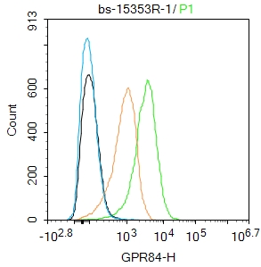
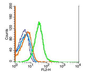
Applications: IHC-P=1:100-500,IHC-F=1:100-500,IF=1:100-500,Flow-Cyt=1μg/Test
Cross Reactive Species: Human,Mouse,Rabbit (predicted: Pig,Cow)
For research use only. Not intended for diagnostic or therapeutic use.