bs-1414R [Primary Antibody]
Rabbit  Anti-HDAC1 Polyclonal Antibody
www.bioss.com.cn
sales@bioss.com.cn
techsupport@bioss.com.cn
400-901-9800
DATASHEET

Host: Rabbit

Target Protein: HDAC1

IR: Immunogen Range:381-482/482

Clonality: Polyclonal

Isotype: IgG

Entrez Gene: 3065

Swiss Prot: Q13547

Source: KLH conjugated synthetic peptide derived from human HDAC1:381-482/482 

Purification: affinity purified by Protein A

Storage: 0.01M TBS(pH7.4) with 1% BSA, 0.03% Proclin300 and 50% Glycerol. Shipped at 4℃. Store at -20 °C for one year. Avoid repeated freeze/thaw cycles.

Background: Histone acetylation and deacetylation, catalyzed by multisubunit complexes, play a key role in the regulation of eukaryotic gene expression. The protein encoded by this gene belongs to the histone deacetylase/acuc/apha family and is a component of the histone deacetylase complex. It also interacts with retinoblastoma tumor-suppressor protein and this complex is a key element in the control of cell proliferation and differentiation. Together with metastasis-associated protein-2, it deacetylates p53 and modulates its effect on cell growth and apoptosis. [provided by RefSeq, Jul 2008]

Size: 50ul

Concentration: 1mg/ml

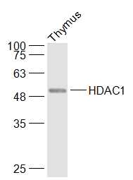
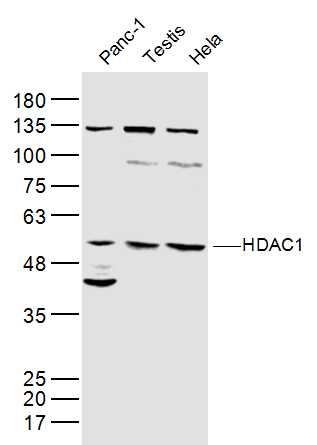
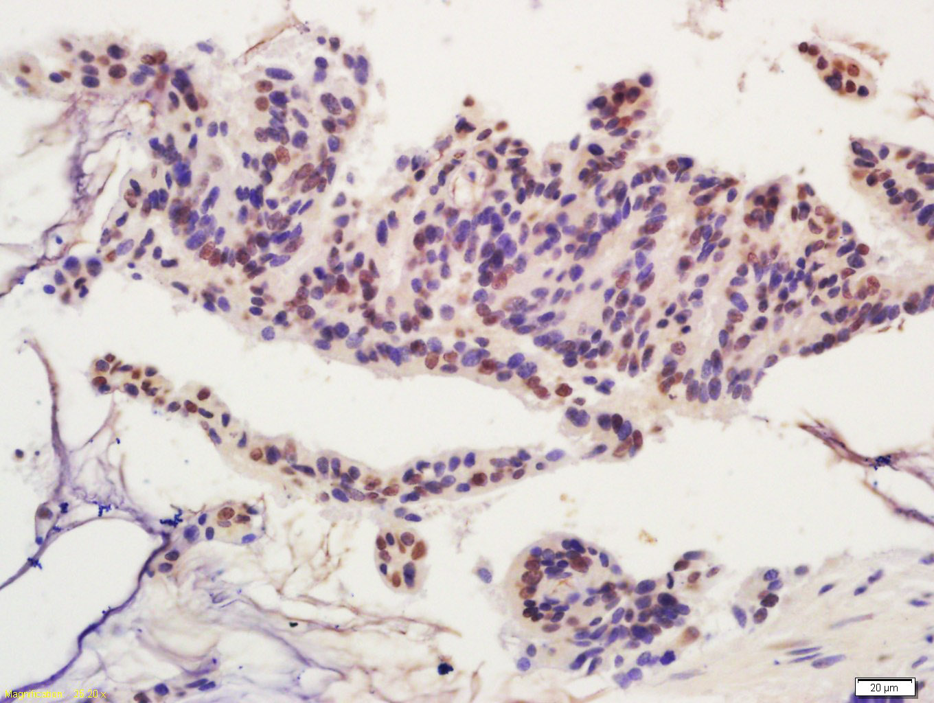
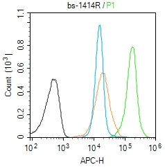
Applications: WB=1:500-2000,IHC-P=1:100-500,IHC-F=1:100-500,Flow-Cyt=1:50-300,IF=1:100-500,ELISA=1:5000-10000

Cross Reactive Species: Human,Mouse,GuineaPig (predicted: Rat,Pig,Sheep,Cow,Dog)

For research use only. Not intended for diagnostic or therapeutic use.

VALIDATION IMAGES
Sample: Thymus (Mouse) Lysate at 40 ug Primary: Anti-HDAC1 (bs-1414R) at 1/1000 dilution Secondary: IRDye800CW Goat Anti-Rabbit IgG at 1/20000 dilution Predicted band size: 53 kD Observed band size: 53 kD
Sample: Panc-1(Human) Cell Lysate at 40 ug Testis (Mouse) Lysate at 40 ug Hela(Human) Lysate at 40 ug Primary: Anti-HDAC1 (bs-1414R) at 1/300 dilution Secondary: IRDye800CW Goat Anti-Rabbit IgG at 1/20000 dilution Predicted band size: 53 kD Observed band size: 53 kD
Tissue/cell: human colon carcinoma; 4% Paraformaldehyde-fixed and paraffin-embedded; Antigen retrieval: citrate buffer ( 0.01M, pH 6.0 ), Boiling bathing for 15min; Block endogenous peroxidase by 3% Hydrogen peroxide for 30min; Blocking buffer (normal goat serum,C-0005) at 37℃ for 20 min; Incubation: Anti-HDAC1 Polyclonal Antibody, Unconjugated(bs-1414R) 1:200, overnight at 4°C, followed by conjugation to the secondary antibody(SP-0023) and DAB(C-0010) staining
Blank control (Black line):Molt4 (Black). Primary Antibody (green line): Rabbit Anti-HDAC1 antibody (bs-1414R) Dilution: 1μg /10^6 cells; Isotype Control Antibody (orange line): Rabbit IgG . Secondary Antibody (white blue line): Goat anti-rabbit IgG-AF647 Dilution: 1μg /test. Protocol The cells were fixed with 4% PFA (10min at room temperature)and then permeabilized with 90% ice-cold methanol for 20 min at room temperature. The cells were then incubated in 5%BSA to block non-specific protein-protein interactions for 30 min at room temperature .Cells stained with Primary Antibody for 30 min at room temperature. The secondary antibody used for 40 min at room temperature. Acquisition of 20,000 events was performed.
 
PRODUCT SPECIFIC PUBLICATIONS