bs-4258R [Primary Antibody]
Rabbit  Anti-Vesicle docking protein p115 Polyclonal Antibody
www.bioss.com.cn
sales@bioss.com.cn
techsupport@bioss.com.cn
400-901-9800
DATASHEET

Host: Rabbit

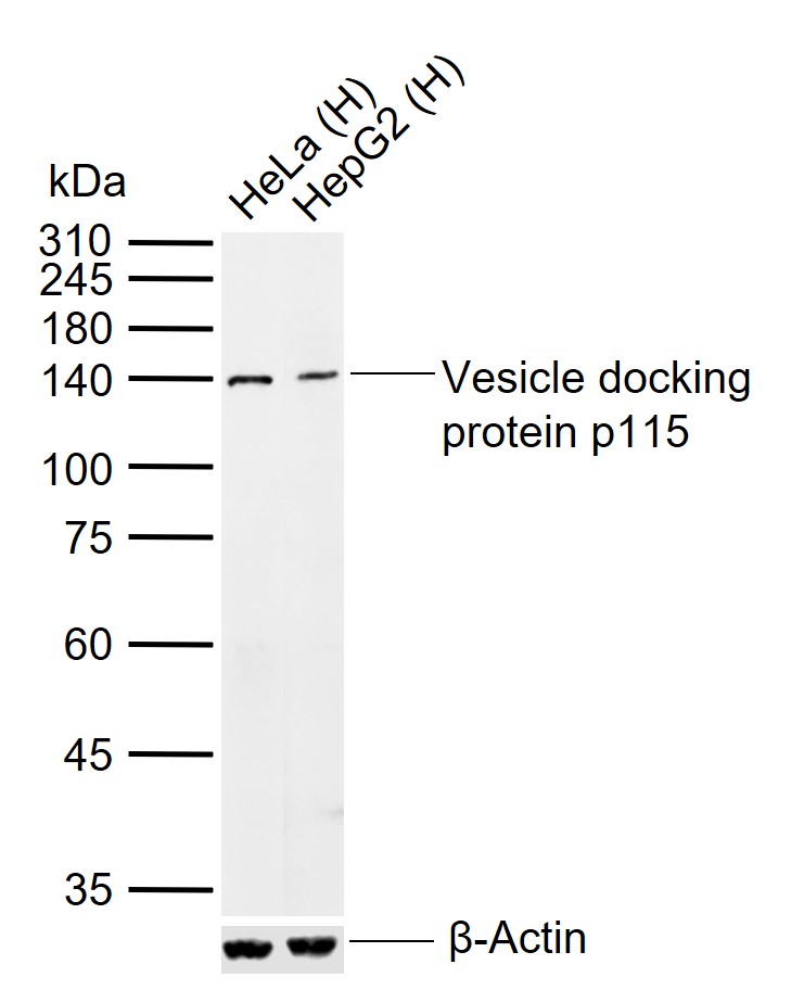
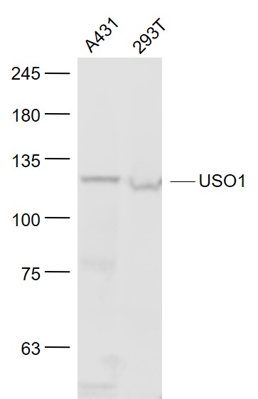
Target Protein: Vesicle docking protein p115

IR: Immunogen Range:501-600/962

Clonality: Polyclonal

Isotype: IgG

Entrez Gene: 8615

Swiss Prot: O60763

Source: KLH conjugated synthetic peptide derived from human Vesicle docking protein p115:501-600/962 

Purification: affinity purified by Protein A

Storage: 0.01M TBS(pH7.4) with 1% BSA, 0.03% Proclin300 and 50% Glycerol. Shipped at 4℃. Store at -20 °C for one year. Avoid repeated freeze/thaw cycles.

Background: p115 (Vesicle docking protein p115) is a peripheral membrane protein that is located on the Golgi complex. p115 exists as a homodimer with two globular heads, an extended coiled-coil tail, followed by an acidic domain at the extreme C terminus. p115 is homologous to a yeast protein, Uso1p, which is required for ER to Golgi transport. p115 likely plays an important role in vesicle transportation from the ER to the cis-Golgi comparments.

Size: 200ul

Concentration: 1mg/ml

Applications: WB=1:500-2000,IHC-P=1:100-500,IHC-F=1:100-500,IF=1:100-500,ELISA=1:5000-10000

Cross Reactive Species: Human,Mouse (predicted: Rat,Pig,Sheep,Cow,Chicken,Horse)

For research use only. Not intended for diagnostic or therapeutic use.

VALIDATION IMAGES
Sample: Lane 1: Human HeLa cell lysates Lane 2: Human HepG2 cell lysates Primary: Anti-Vesicle docking protein p115 (bs-4258R) at 1/1000 dilution Secondary: IRDye800CW Goat Anti-Rabbit IgG at 1/20000 dilution Predicted band size: 108 kDa Observed band size: 140 kDa
Sample: A431(Human) Cell Lysate at 30 ug 293T(Human) Cell Lysate at 30 ug Primary: Anti- USO1 (bs-4258R) at 1/1000 dilution Secondary: IRDye800CW Goat Anti-Rabbit IgG at 1/20000 dilution Predicted band size: 108 kD Observed band size: 120 kD
Paraformaldehyde-fixed, paraffin embedded (Mouse brain); Antigen retrieval by boiling in sodium citrate buffer (pH6.0) for 15min; Block endogenous peroxidase by 3% hydrogen peroxide for 20 minutes; Blocking buffer (normal goat serum) at 37°C for 30min; Antibody incubation with (Vesicle docking protein p115) Polyclonal Antibody, Unconjugated (bs-4258R) at 1:400 overnight at 4°C, followed by operating according to SP Kit(Rabbit) (sp-0023) instructionsand DAB staining.
Paraformaldehyde-fixed, paraffin embedded (mouse brain tissue); Antigen retrieval by boiling in sodium citrate buffer (pH6.0) for 15min; Block endogenous peroxidase by 3% hydrogen peroxide for 20 minutes; Blocking buffer (normal goat serum) at 37°C for 30min; Antibody incubation with (Vesicle docking protein p115) Polyclonal Antibody, Unconjugated (bs-4258R) at 1:400 overnight at 4°C, followed by a conjugated secondary (sp-0023) for 20 minutes and DAB staining.
 
PRODUCT SPECIFIC PUBLICATIONS