sales@bioss.com.cn
techsupport@bioss.com.cn
400-901-9800
Host: Rabbit
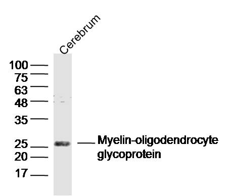
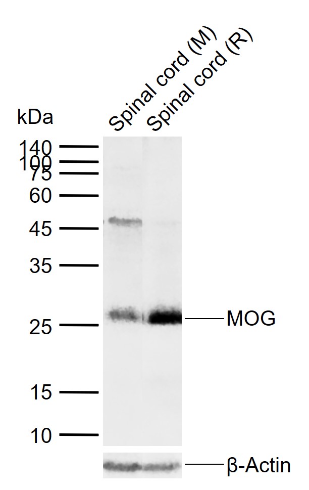
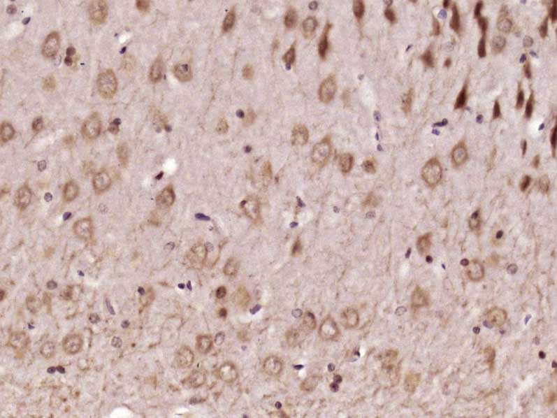
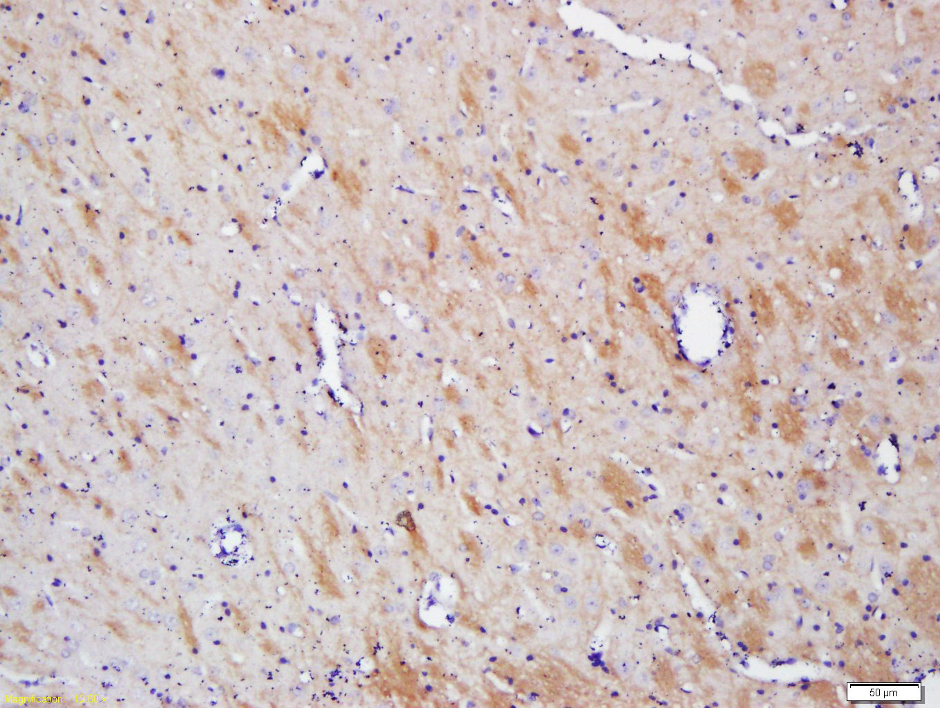
Target Protein: MOG
IR: Immunogen Range:35-55/247
Clonality: Polyclonal
Isotype: IgG
Entrez Gene: 4340
Swiss Prot: Q61885
Source:
KLH conjugated synthetic peptide derived from mouse MOG:35-55/247
Purification: affinity purified by Protein A
Storage: 0.01M TBS(pH7.4) with 1% BSA, 0.03% Proclin300 and 50% Glycerol. Shipped at 4℃. Store at -20 °C for one year. Avoid repeated freeze/thaw cycles.
Background: The product of this gene is a membrane protein expressed on the oligodendrocyte cell surface and the outermost surface of myelin sheaths. Due to this localization, it is a primary target antigen involved in immune-mediated demyelination. This protein may be involved in completion and maintenance of the myelin sheath and in cell-cell communication. Alternatively spliced transcript variants encoding different isoforms have been identified. [provided by RefSeq, Jul 2008]
Size: 200ul
Concentration: 1mg/ml
Applications: WB=1:500-2000,IHC-P=1:100-500,IHC-F=1:100-500,ICC=1:100-500,IF=1:100-500,ELISA=1:5000-10000
Cross Reactive Species: Mouse,Rat (predicted: Human,Pig,GuineaPig)
For research use only. Not intended for diagnostic or therapeutic use.