bs-7052R [Primary Antibody]
Rabbit  Anti-Myosin light chain (phospho S20)  Polyclonal Antibody
www.bioss.com.cn
sales@bioss.com.cn
techsupport@bioss.com.cn
400-901-9800
DATASHEET

Host: Rabbit

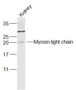
Target Protein: Myosin light chain (phospho S20)

IR: Immunogen Range:AT(p-S)NV

Clonality: Polyclonal

Isotype: IgG

Entrez Gene: 10398

Swiss Prot: P24844

Source: KLH conjugated synthesised phosphopeptide derived from human MYL9 around the phosphorylation site of Ser20:AT(p-S)NV 

Purification: affinity purified by Protein A

Storage: 0.01M TBS(pH7.4) with 1% BSA, 0.03% Proclin300 and 50% Glycerol. Shipped at 4℃. Store at -20 °C for one year. Avoid repeated freeze/thaw cycles.

Background: Myosin light chain (MLC) is a subunit of the conventional myosins (e.g. myosin II). In smooth muscle and non-muscle cells conventional myosins mediate a wide variety of contractile events including cytokinesis, cell motility, and smooth muscle contraction. MLC is phosphorylated by multiple serine-threonine kinases such as Rho-kinase and PAK, however myosin light chain kinase (MLCK) acts as the primary kinase. Contractile activity of conventional myosins is regulated by phosphorylation of MLC on several residues.

Size: 200ul

Concentration: 1mg/ml

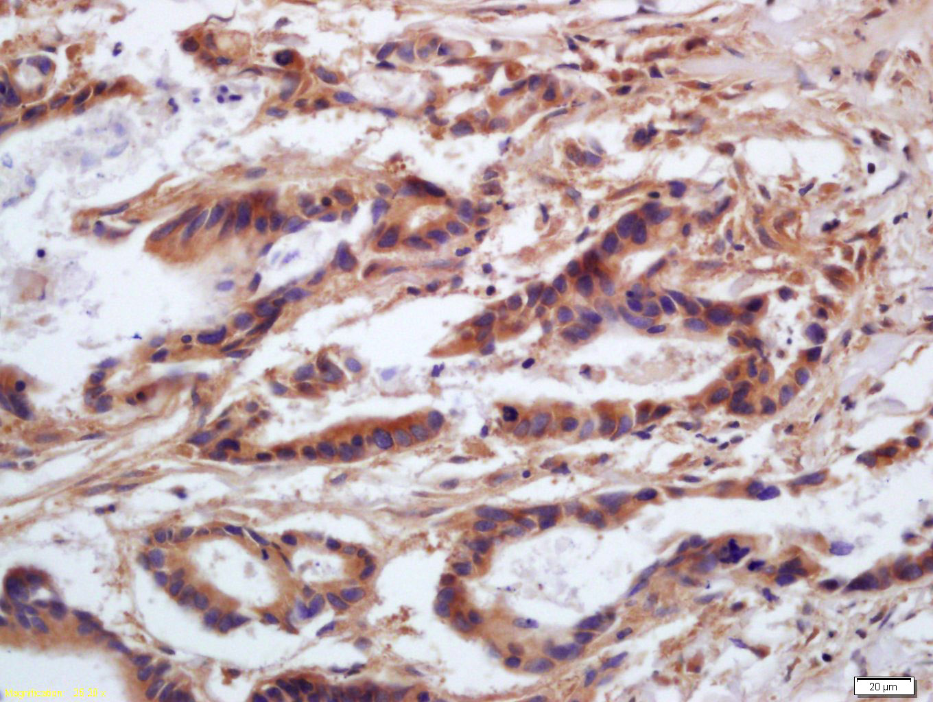
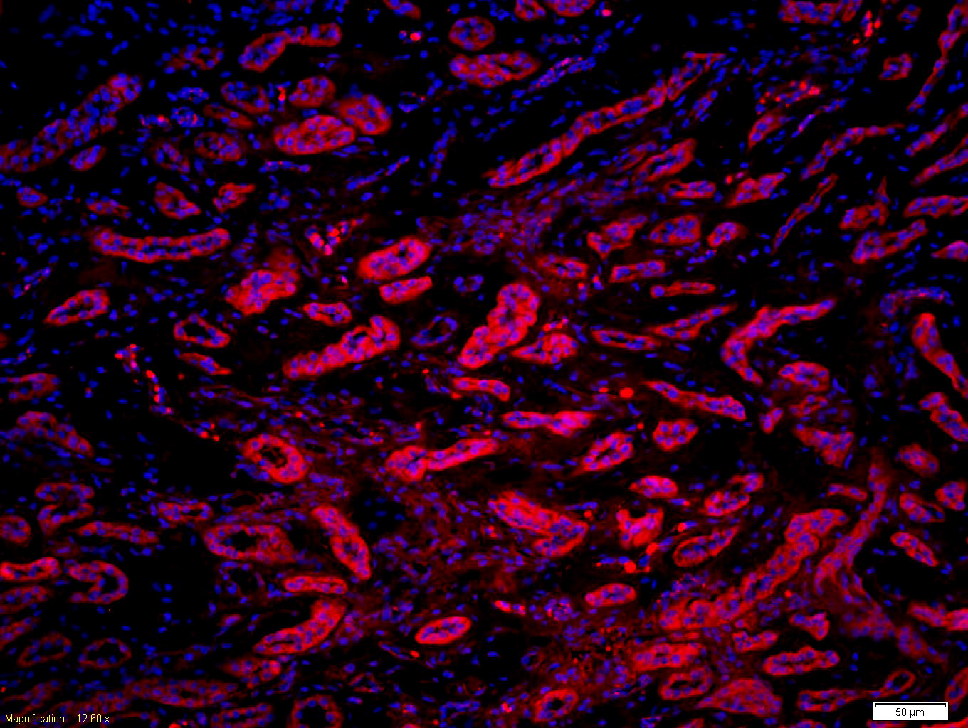
Applications: WB=1:500-2000,IHC-P=1:100-500,IHC-F=1:100-500,IF=1:100-500,ELISA=1:5000-10000

Cross Reactive Species: Human (predicted: Mouse,Rat,Rabbit,Pig,Sheep,Cow,Dog)

For research use only. Not intended for diagnostic or therapeutic use.

VALIDATION IMAGES
Sample: Kidney (Mouse) Lysate at 40 ug Primary: Anti-Myosin light chain (phospho S20) (bs-7052R) at 1/1000 dilution Secondary: IRDye800CW Goat Anti-Rabbit IgG at 1/20000 dilution Predicted band size: 20 kD Observed band size: 20 kD
Tissue/cell: human gastric carcinoma; 4% Paraformaldehyde-fixed and paraffin-embedded; Antigen retrieval: citrate buffer ( 0.01M, pH 6.0 ), Boiling bathing for 15min; Block endogenous peroxidase by 3% Hydrogen peroxide for 30min; Blocking buffer (normal goat serum,C-0005) at 37℃ for 20 min; Incubation: Anti-phospho-MLC(Ser20) Polyclonal Antibody, Unconjugated(bs-7052R) 1:200, overnight at 4°C, followed by conjugation to the secondary antibody(SP-0023) and DAB(C-0010) staining
Tissue/cell: human kidney tissue; 4% Paraformaldehyde-fixed and paraffin-embedded; Antigen retrieval: citrate buffer ( 0.01M, pH 6.0 ), Boiling bathing for 15min; Block endogenous peroxidase by 3% Hydrogen peroxide for 30min; Blocking buffer (normal goat serum,C-0005) at 37℃ for 20 min; Incubation: Anti-phospho-MLC(Ser20) Polyclonal Antibody, Unconjugated(bs-7052R) 1:200, overnight at 4°C, followed by conjugation to the secondary antibody(SP-0023) and DAB(C-0010) staining
Tissue/cell: human kidney tissue;4% Paraformaldehyde-fixed and paraffin-embedded; Antigen retrieval: citrate buffer ( 0.01M, pH 6.0 ), Boiling bathing for 15min; Blocking buffer (normal goat serum,C-0005) at 37℃ for 20 min; Incubation: Anti-phospho-MLC(Ser20) Polyclonal Antibody, Unconjugated(bs-7052R) 1:200, overnight at 4°C; The secondary antibody was Goat Anti-Rabbit IgG, Cy3 conjugated (bs-0295G-Cy3)used at 1:200 dilution for 40 minutes at 37°C. DAPI(5ug/ml,blue,C-0033) was used to stain the cell nuclei
 
PRODUCT SPECIFIC PUBLICATIONS