bs-0559R [Primary Antibody]
Rabbit  Anti-CDK5  Polyclonal Antibody
www.bioss.com.cn
sales@bioss.com.cn
techsupport@bioss.com.cn
400-901-9800
DATASHEET

Host: Rabbit

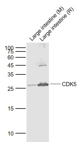
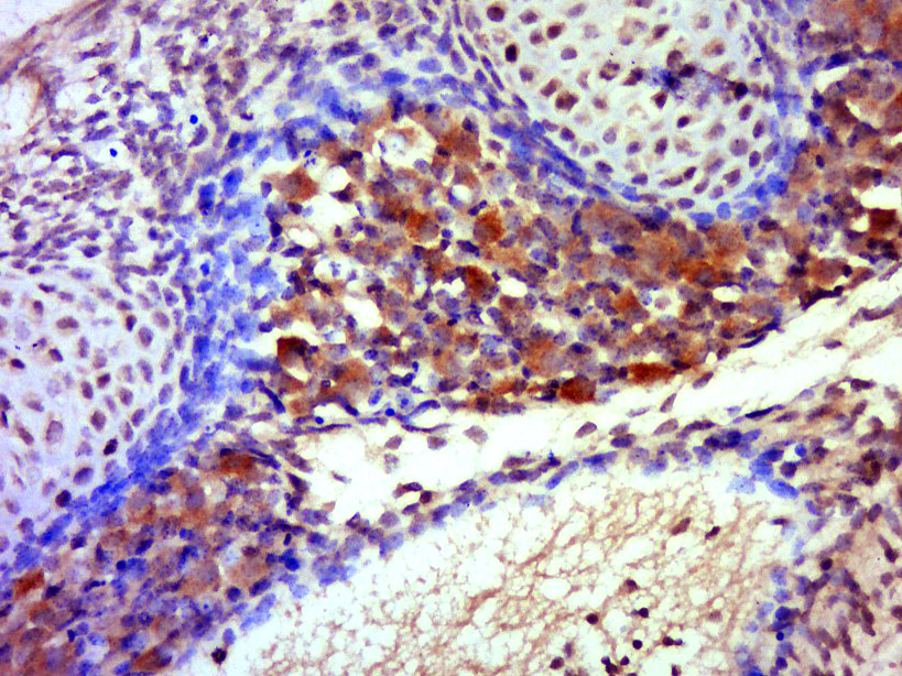
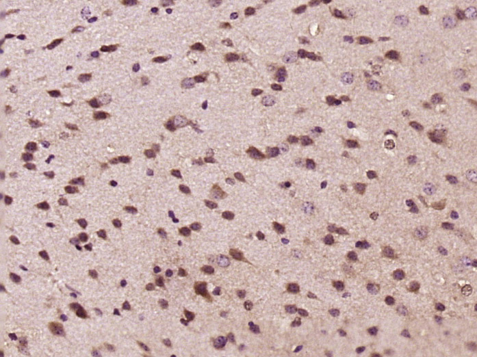
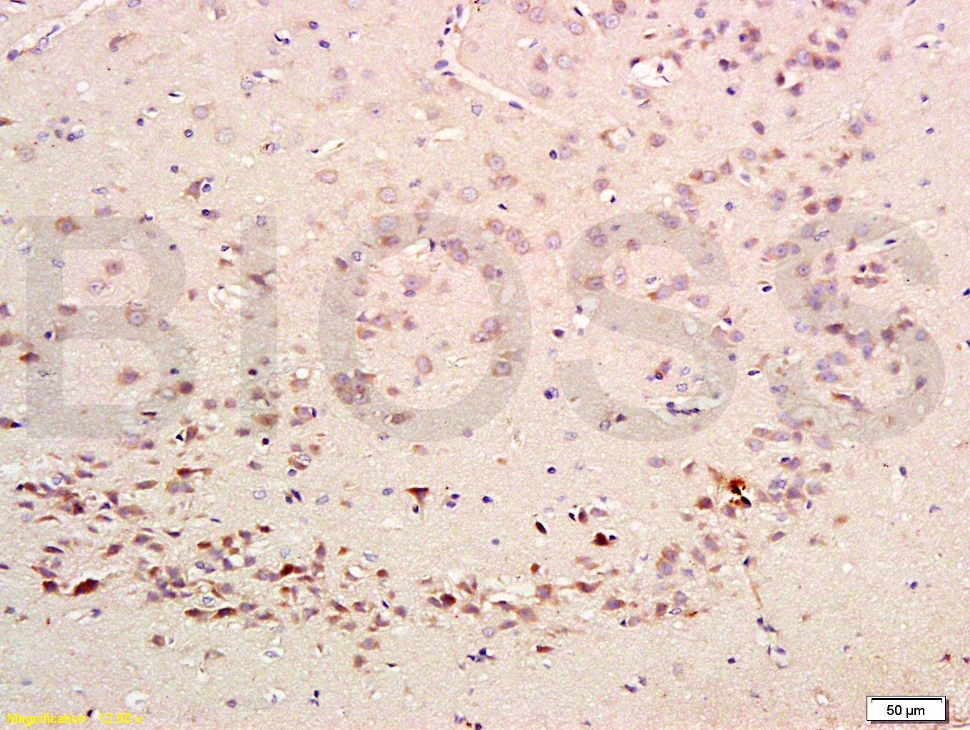
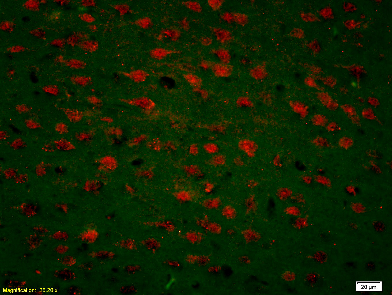
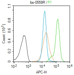
Target Protein: CDK5

IR: Immunogen Range:15-100/292

Clonality: Polyclonal

Isotype: IgG

Entrez Gene: 1020

Swiss Prot: Q00535

Source: KLH conjugated synthetic peptide derived from human CDK5:15-100/292 

Purification: affinity purified by Protein A

Storage: 0.01M TBS (pH7.4) with 1% BSA, 0.02% Proclin300 and 50% Glycerol. Shipped at 4℃. Store at -20℃ for one year. Avoid repeated freeze/thaw cycles.

Background: This gene encodes a proline-directed serine/threonine kinase that is a member of the cyclin-dependent kinase family of proteins. Unlike other members of the family, the protein encoded by this gene does not directly control cell cycle regulation. Instead the protein, which is predominantly expressed at high levels in mammalian postmitotic central nervous system neurons, functions in diverse processes such as synaptic plasticity and neuronal migration through phosphorylation of proteins required for cytoskeletal organization, endocytosis and exocytosis, and apoptosis. In humans, an allelic variant of the gene that results in undetectable levels of the protein has been associated with lethal autosomal recessive lissencephaly-7. Alternative splicing results in multiple transcript variants. [provided by RefSeq, May 2015]

Size: 200ul

Concentration: 1mg/ml

Applications: WB=1:500-2000,IHC-P=1:100-500,IHC-F=1:100-500,Flow-Cyt=1ug/Test,IF=1:100-500

Cross Reactive Species: Human,Mouse,Rat (predicted: Pig,Cow)

For research use only. Not intended for diagnostic or therapeutic use.

VALIDATION IMAGES
Sample: Lane 1: Large intestine (Mouse) Lysate at 40 ug Lane 2: Large intestine (Rat) Lysate at 40 ug Primary: Anti-CDK5 (bs-0559R) at 1/1000 dilution Secondary: IRDye800CW Goat Anti-Rabbit IgG at 1/20000 dilution Predicted band size: 29 kD Observed band size: 27 kD
Tissue/cell: mouse embryo tissue; 4% Paraformaldehyde-fixed and paraffin-embedded; Antigen retrieval: citrate buffer ( 0.01M, pH 6.0 ), Boiling bathing for 15min; Block endogenous peroxidase by 3% Hydrogen peroxide for 30min; Blocking buffer (normal goat serum,C-0005) at 37℃ for 20 min; Incubation: Anti-CDK5 Polyclonal Antibody, Unconjugated(bs-0559R) 1:500, overnight at 4°C, followed by conjugation to the secondary antibody(SP-0023) and DAB(C-0010) staining
Paraformaldehyde-fixed, paraffin embedded (mouse brain tissue); Antigen retrieval by boiling in sodium citrate buffer (pH6.0) for 15min; Block endogenous peroxidase by 3% hydrogen peroxide for 20 minutes; Blocking buffer (normal goat serum) at 37°C for 30min; Antibody incubation with (CDK5) Polyclonal Antibody, Unconjugated (bs-0559R) at 1:400 overnight at 4°C, followed by operating according to SP Kit(Rabbit) (sp-0023) instructionsand DAB staining.
Tissue/cell: rat brain tissue; 4% Paraformaldehyde-fixed and paraffin-embedded; Antigen retrieval: citrate buffer ( 0.01M, pH 6.0 ), Boiling bathing for 15min; Block endogenous peroxidase by 3% Hydrogen peroxide for 30min; Blocking buffer (normal goat serum,C-0005) at 37℃ for 20 min; Incubation: Anti-CDK5 Polyclonal Antibody, Unconjugated(bs-0559R) 1:300, overnight at 4°C, followed by conjugation to the secondary antibody(SP-0023) and DAB(C-0010) staining
Tissue/cell: rat brain tissue;4% Paraformaldehyde-fixed and paraffin-embedded; Antigen retrieval: citrate buffer ( 0.01M, pH 6.0 ), Boiling bathing for 15min; Blocking buffer (normal goat serum,C-0005) at 37℃ for 20 min; Incubation: Anti-CDK5 Polyclonal Antibody, Unconjugated(bs-0559R) 1:200, overnight at 4°C; The secondary antibody was Goat Anti-Rabbit IgG, Cy3 conjugated(bs-0295G-Cy3)used at 1:200 dilution for 40 minutes at 37°C.
Blank control (Black line):Molt4 (Black). Primary Antibody (green line): Rabbit Anti-CDK5 antibody (bs-0559R) Dilution: 1μg /10^6 cells; Isotype Control Antibody (orange line): Rabbit IgG . Secondary Antibody (white blue line): Goat anti-rabbit IgG-AF647 Dilution: 1μg /test. Protocol The cells were fixed with 4% PFA (10min at room temperature)and then permeabilized with 90% ice-cold methanol for 20 min at room temperature. The cells were then incubated in 5%BSA to block non-specific protein-protein interactions for 30 min at room temperature .Cells stained with Primary Antibody for 30 min at room temperature. The secondary antibody used for 40 min at room temperature. Acquisition of 20,000 events was performed.
 
PRODUCT SPECIFIC PUBLICATIONS