bs-12048R [Primary Antibody]
Rabbit  Anti-5HT1F Receptor  Polyclonal Antibody
www.bioss.com.cn
sales@bioss.com.cn
techsupport@bioss.com.cn
400-901-9800
DATASHEET

Host: Rabbit

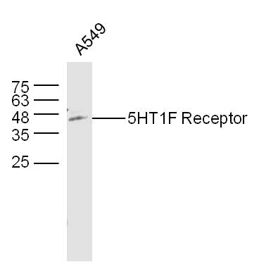
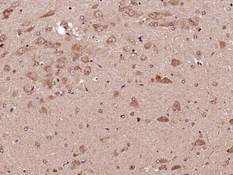
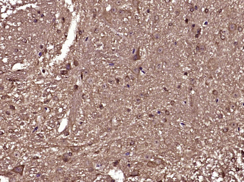
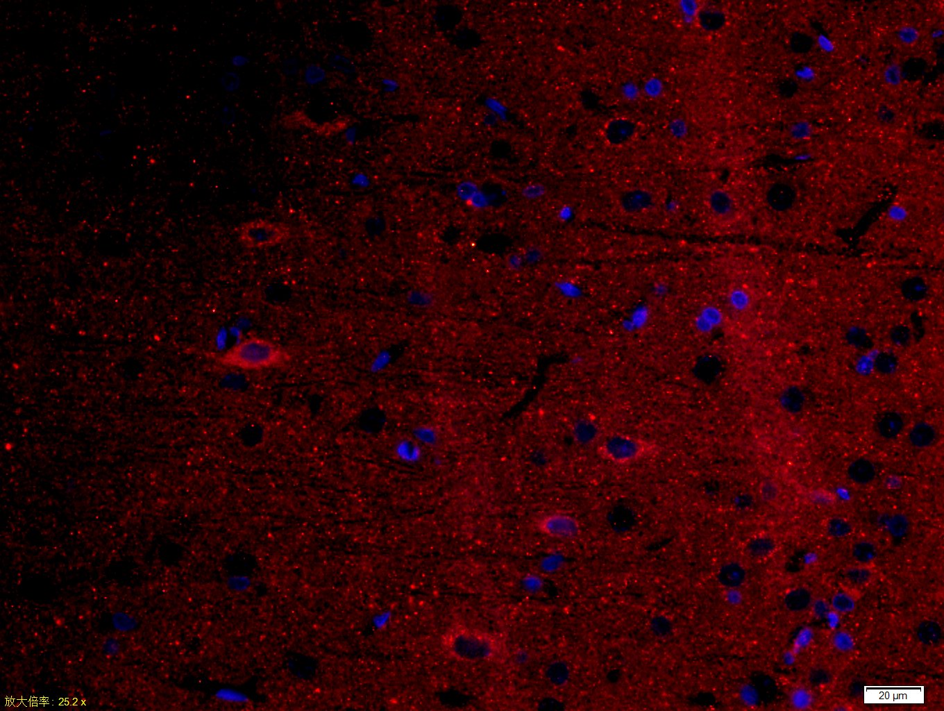
Target Protein: 5HT1F Receptor

IR: Immunogen Range:1-100/366

Clonality: Polyclonal

Isotype: IgG

Entrez Gene: 3355

Swiss Prot: P30939

Source: KLH conjugated synthetic peptide derived from human 5HT1F Receptor/SR-1F:1-100/366 

Purification: affinity purified by Protein A

Storage: 0.01M TBS (pH7.4) with 1% BSA, 0.02% Proclin300 and 50% Glycerol. Shipped at 4℃. Store at -20℃ for one year. Avoid repeated freeze/thaw cycles.

Background: The members of the G-protein-coupled receptor family are distinguished by their slow transmitting response to ligand binding. These seven transmembrane proteins include the adrenergic, serotonin and dopamine receptors. The effect of the signaling molecule can be excitatory or inhibitory depending on the type of receptor to which it binds. b-adrenergic bound to adrenaline activates adenylyl cyclase, while a2-adrenergic receptor bound to adrenaline inhibits adenylyl cyclase. Like the a2-adrenergic receptor, serotonin receptor functions are also mediated by G proteins that inhibit the activity of adenylyl cyclase. The serotonin receptors have been classified into several categories, designated SR-1–7 (5HT1–7). Subtypes within the SR-1 group include SR-1A, -1B, -1D, -1E and -1F.

Size: 200ul

Concentration: 1mg/ml

Applications: WB=1:500-2000,IHC-P=1:100-500,IHC-F=1:100-500,ICC/IF=1:100-500,IF=1:100-500,ELISA=1:5000-10000

Cross Reactive Species: Human,Mouse,Rat (predicted: Rabbit,Pig,Sheep,Cow,Dog)

For research use only. Not intended for diagnostic or therapeutic use.

VALIDATION IMAGES
Sample: A549 Cell (Human) Lysate at 40 ug Primary: Anti- 5HT1F Receptor (bs-12048R) at 1/300 dilution Secondary: IRDye800CW Goat Anti-Rabbit IgG at 1/20000 dilution Predicted band size: 42 kD Observed band size: 42 kD
Sample: Lane 1: Mouse Cerebrum tissue lysates Lane 2: Rat Cerebrum tissue lysates Lane 3: Rat Eye tissue lysates Primary: Anti-5HT1F Receptor (bs-12048R) at 1/1000 dilution Secondary: IRDye800CW Goat Anti-Rabbit IgG at 1/20000 dilution Predicted band size: 42 kDa Observed band size: 48 kDa
Paraformaldehyde-fixed, paraffin embedded (Mouse brain); Antigen retrieval by boiling in sodium citrate buffer (pH6.0) for 15min; Block endogenous peroxidase by 3% hydrogen peroxide for 20 minutes; Blocking buffer (normal goat serum) at 37°C for 30min; Antibody incubation with (5HT1F Receptor) Polyclonal Antibody, Unconjugated (bs-12048R) at 1:400 overnight at 4°C, followed by operating according to SP Kit(Rabbit) (sp-0023) instructionsand DAB staining.
Paraformaldehyde-fixed, paraffin embedded (Mouse spinal cord); Antigen retrieval by boiling in sodium citrate buffer (pH6.0) for 15min; Block endogenous peroxidase by 3% hydrogen peroxide for 20 minutes; Blocking buffer (normal goat serum) at 37°C for 30min; Antibody incubation with (5HT1F Receptor) Polyclonal Antibody, Unconjugated (bs-12048R) at 1:400 overnight at 4°C, followed by operating according to SP Kit(Rabbit) (sp-0023) instructionsand DAB staining.
Tissue/cell: rat brain tissue; 4% Paraformaldehyde-fixed and paraffin-embedded; Antigen retrieval: citrate buffer ( 0.01M, pH 6.0 ), Boiling bathing for 15min; Block endogenous peroxidase by 3% Hydrogen peroxide for 30min; Blocking buffer (normal goat serum,C-0005) at 37℃ for 20 min; Incubation: Anti-5HT1F Polyclonal Antibody, Unconjugated(bs-12048R) 1:500, overnight at 4°C, followed by conjugation to the secondary antibody(SP-0023) and DAB(C-0010) staining
Tissue/cell: rat brain tissue;4% Paraformaldehyde-fixed and paraffin-embedded; Antigen retrieval: citrate buffer ( 0.01M, pH 6.0 ), Boiling bathing for 15min; Blocking buffer (normal goat serum,C-0005) at 37℃ for 20 min; Incubation: Anti-5HT1F Receptor Polyclonal Antibody, Unconjugated(bs-12048R) 1:200, overnight at 4°C; The secondary antibody was Goat Anti-Rabbit IgG, Cy3 conjugated(bs-0295G-Cy3)used at 1:200 dilution for 40 minutes at 37°C. DAPI(5ug/ml,blue,C-0033) was used to stain the cell nuclei
 
PRODUCT SPECIFIC PUBLICATIONS