bs-1306R [Primary Antibody]
Rabbit  Anti-PARK7/DJ1  Polyclonal Antibody
www.bioss.com.cn
sales@bioss.com.cn
techsupport@bioss.com.cn
400-901-9800
DATASHEET

Host: Rabbit

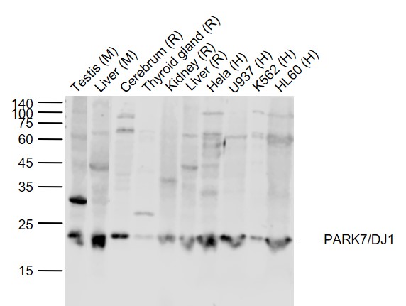
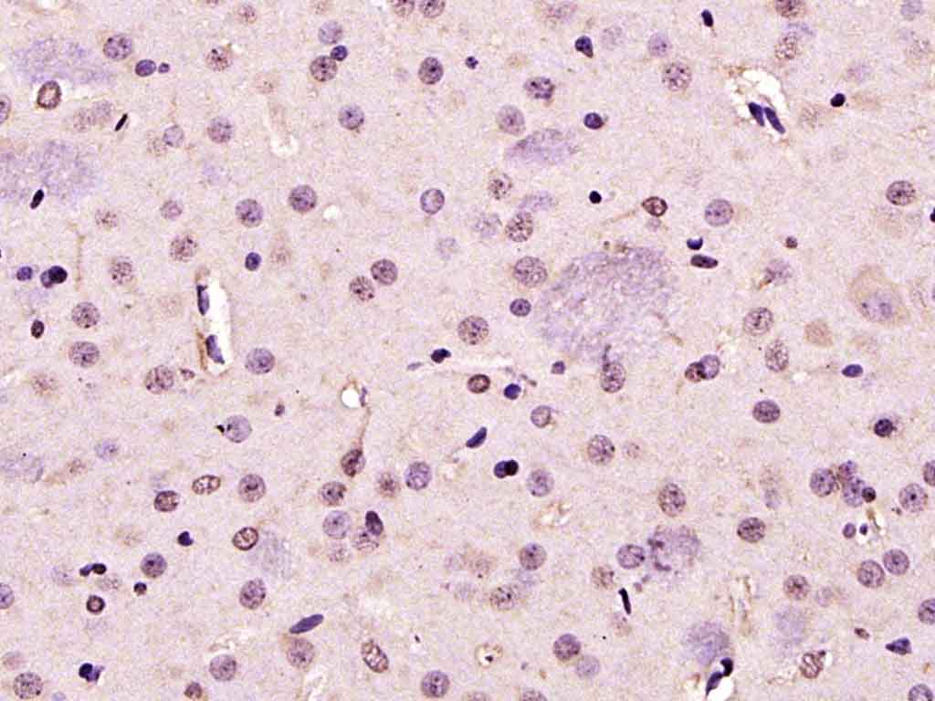
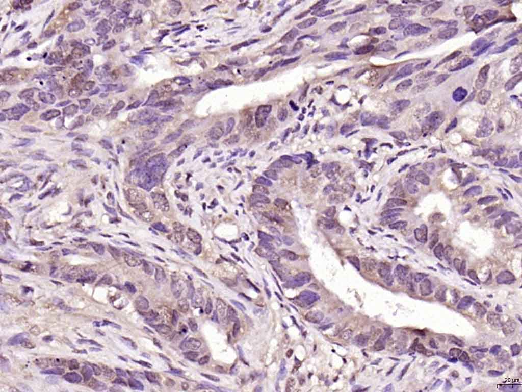
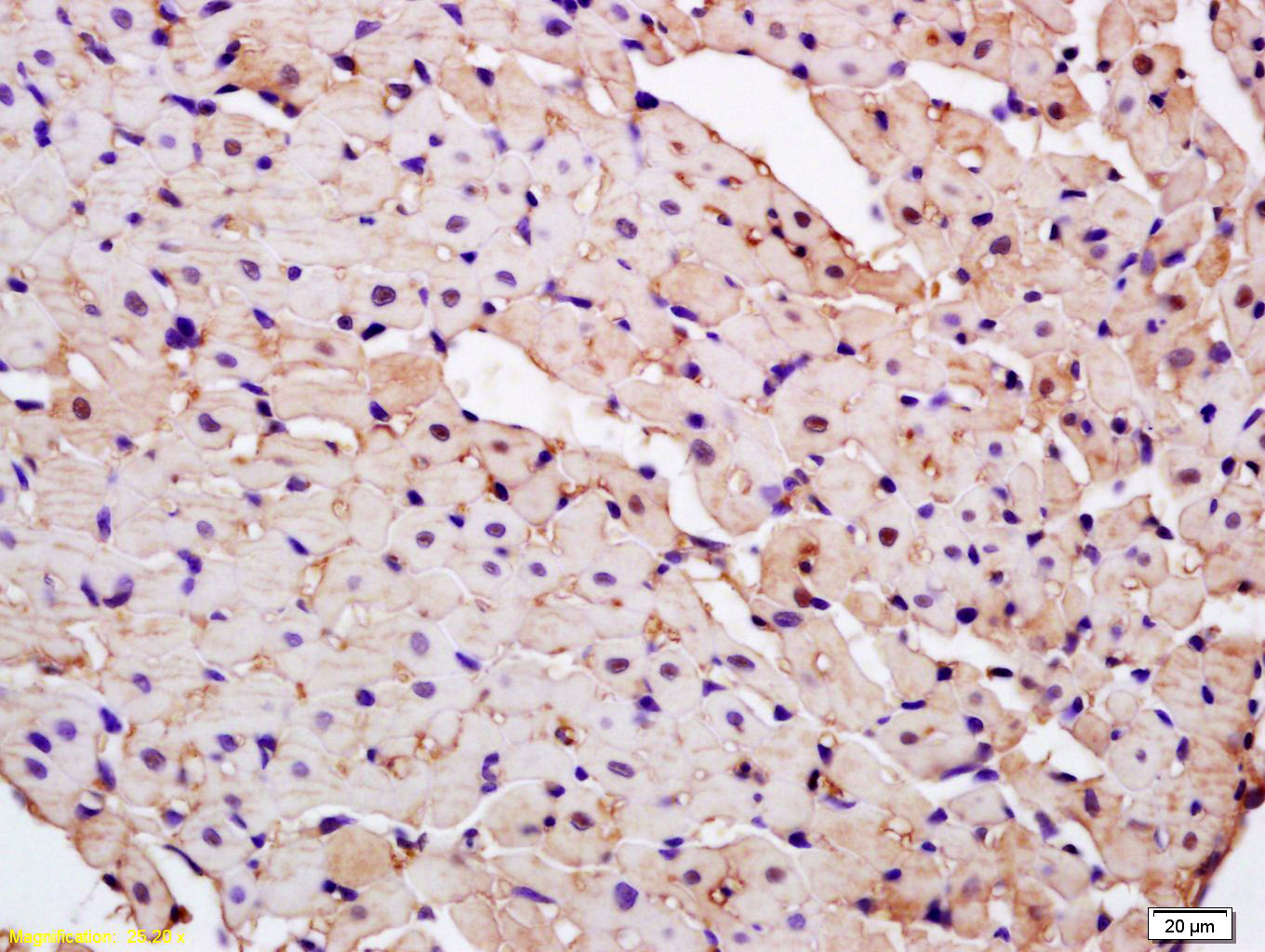
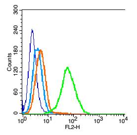
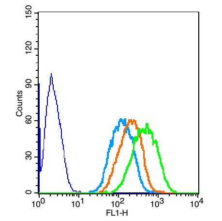
Target Protein: PARK7/DJ1

IR: Immunogen Range:101-189/189

Clonality: Polyclonal

Isotype: IgG

Entrez Gene: 11315

Swiss Prot: Q99497

Source: KLH conjugated synthetic peptide derived from human CAP1:101-189/189 

Purification: affinity purified by Protein A

Storage: 0.01M TBS (pH7.4) with 1% BSA, 0.02% Proclin300 and 50% Glycerol. Shipped at 4℃. Store at -20℃ for one year. Avoid repeated freeze/thaw cycles.

Background: PARK7/DJ1 is a ubiquitously expressed protein involved in various cellular processes including cell proliferation, RNA-binding, and oxidative stress. The protein has been found to colocalize within a subset of pathologic tau inclusions in a diverse group of neurodegenerative disorders known as tauopathies (Rizzu et al. 2004). Defects in PARK7/DJ1 are the cause of autosomal recessive early-onset Parkinson's disease 7 (PARK7). Parkinson's disease (PD) is a complex, multifactorial disorder that typically manifests after the age of 50 years. The disease is characterized by bradykinesia, resting tremor, muscular rigidity and postural instability. The pathology involves the loss of dopaminergic neurons in the substantia nigra and the presence of Lewy bodies (intraneuronal accumulations of aggregated proteins), in surviving neurons in various areas of the brain. PARK7 is characterized by onset before 40 years and slow progression. It has also been suggested that PARK7/DJ1 is a mitogen dependent oncogene product involved in Ras related signal transduction pathways.

Size: 200ul

Concentration: 1mg/ml

Applications: WB=1:500-2000,IHC-P=1:100-500,IHC-F=1:100-500,Flow-Cyt=0.2μg/Test,IF=1:100-500,ELISA=1:5000-10000

Cross Reactive Species: Human,Mouse,Rat (predicted: Pig,Cow,Horse)

For research use only. Not intended for diagnostic or therapeutic use.

VALIDATION IMAGES
Sample: Lane 1: Testis (Mouse) Lysate at 40 ug Lane 2: Liver (Mouse) Lysate at 40 ug Lane 3: Cerebrum (Rat) Lysate at 40 ug Lane 4: Thyroid gland Rat) Lysate at 40 ug Lane 5: Kidney (Rat) Lysate at 40 ug Lane 6: Liver (Rat) Lysate at 40 ug Lane 7: Hela (Human) Cell Lysate at 30 ug Lane 8: U937 (Human) Cell Lysate at 30 ug Lane 9: K562 (Human) Cell Lysate at 30 ug Lane 10: HL60 (Human) Cell Lysate at 30 ug Primary: Anti-PARK7/DJ1 (bs-1306R) at 1/1000 dilution Secondary: IRDye800CW Goat Anti-Rabbit IgG at 1/20000 dilution Predicted band size: 22 kD Observed band size: 22 kD
Paraformaldehyde-fixed, paraffin embedded (rat brain); Antigen retrieval by boiling in sodium citrate buffer (pH6.0) for 15min; Block endogenous peroxidase by 3% hydrogen peroxide for 20 minutes; Blocking buffer (normal goat serum) at 37°C for 30min; Antibody incubation with (PARK7) Polyclonal Antibody, Unconjugated (bs-1306R ) at 1:200 overnight at 4°C, followed by operating according to SP Kit(Rabbit) (sp-0023) instructionsand DAB staining.
Paraformaldehyde-fixed, paraffin embedded (human colon carcinoma); Antigen retrieval by boiling in sodium citrate buffer (pH6.0) for 15min; Block endogenous peroxidase by 3% hydrogen peroxide for 20 minutes; Blocking buffer (normal goat serum) at 37°C for 30min; Antibody incubation with (PARK7) Polyclonal Antibody, Unconjugated (bs-1306R ) at 1:200 overnight at 4°C, followed by operating according to SP Kit(Rabbit) (sp-0023) instructionsand DAB staining.
Tissue/cell: rat kidney tissue; 4% Paraformaldehyde-fixed and paraffin-embedded; Antigen retrieval: citrate buffer ( 0.01M, pH 6.0 ), Boiling bathing for 15min; Block endogenous peroxidase by 3% Hydrogen peroxide for 30min; Blocking buffer (normal goat serum,C-0005) at 37℃ for 20 min; Incubation: Anti-CAP1/PARK7 Polyclonal Antibody, Unconjugated(bs-1306R) 1:200, overnight at 4°C, followed by conjugation to the secondary antibody(SP-0023) and DAB(C-0010) staining
Blank control: 293T cells(blue). Primary Antibody: Rabbit Anti-PARK7/CAP1 antibody(bs-1306R), Dilution: 0.2μg in 100 μL 1X PBS containing 0.5% BSA; Isotype Control Antibody: Rabbit IgG (orange) ,used under the same conditions. Secondary Antibody: Goat anti-rabbit IgG-PE(white blue), Dilution: 1:200 in 1 X PBS containing 0.5% BSA. Protocol Primary antibody (bs-1306R, 0.2μg /1x10^6 cells) were incubated for 30 min on the ice, followed by 1 X PBS containing 0.5% BSA + 1 0% goat serum (15 min) to block non-specific protein-protein interactions. Then the Goat Anti-rabbit IgG/PE antibody was added into the blocking buffer mentioned above to react with the primary antibody at 1/200 dilution for 30 min on ice. Acquisition of 20,000 events was performed.
The blue histogram is unstained cells (Hepg2 cells) concentration 1:50 The Wathet Blue histogram is cells stained with secondary antibody alone. The Orange histogram is cells stained with rabbit IgG isotype control antibody plus secondary antibody. The green histogram is cells stained with Rabbit Anti-PARK7/CAP1 antibody (bs-1306R)plus secondary antibody.
 
PRODUCT SPECIFIC PUBLICATIONS