bs-0061R [Primary Antibody]
Rabbit  Anti-beta-Actin (Loading Control) Polyclonal Antibody
www.bioss.com.cn
sales@bioss.com.cn
techsupport@bioss.com.cn
400-901-9800
DATASHEET

Host: Rabbit

Target Protein: beta-Actin (Loading Control)

IR: Immunogen Range:1-200/375

Clonality: Polyclonal

Isotype: IgG

Entrez Gene: 60

Swiss Prot: P60709

Source: Synthetic MAP peptide derived from human beta-Actin:1-200/375 

Purification: affinity purified by Protein A

Storage: 0.01M TBS(pH7.4) with 1% BSA, 0.03% Proclin300 and 50% Glycerol. Shipped at 4℃. Store at -20 °C for one year. Avoid repeated freeze/thaw cycles.

Background: Loading Control
This gene encodes one of six different actin proteins. Actins are highly conserved proteins that are involved in cell motility, structure, and integrity. This actin is a major constituent of the contractile apparatus and one of the two nonmuscle cytoskeletal actins. [provided by RefSeq, Jul 2008].

Size: 100ul

Concentration: 1mg/ml

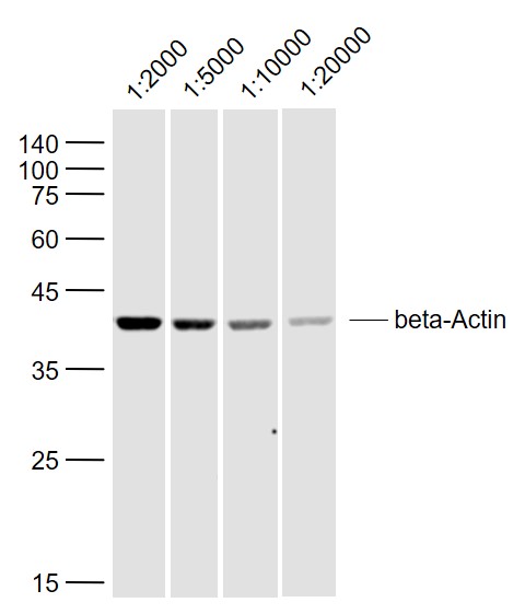
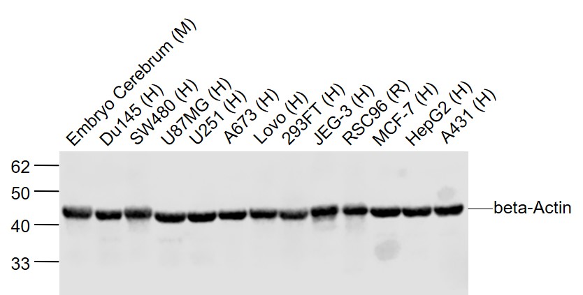
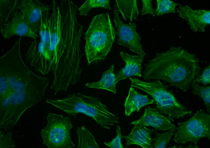
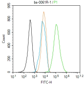
Applications: WB=1:5000-50000,Flow-Cyt=1μg/Test,ICC=1:100,ELISA=1:5000-20000

Cross Reactive Species: Human,Mouse,Rat,Hamster (predicted: Rabbit,Pig,Sheep,Chicken,Dog,Cat,GuineaPig,Fish,Bee)

For research use only. Not intended for diagnostic or therapeutic use.

VALIDATION IMAGES
Sample: SH-SY5Y (Human) Lysate at 40 ug Primary: Anti-beta-Actin (bs-0061R) at 1/2000~1/20000 dilution Secondary: IRDye800CW Goat Anti-Rabbit IgG at 1/20000 dilution Predicted band size: 42 kD Observed band size: 42 kD
Sample:Embryo Cerebrum (Mouse) Lysate at 40 ugDu145 (Human) Lysate at 40 ugSW480 (Human) Cell Lysate at 40 ugU87MG (Human) Lysate at 40 ugU251 (Human) Lysate at 40 ugA673 (Human) Lysate at 40 ugLovo (Human) Lysate at 40 ug293FT (Human) Lysate at 40 ugJEG-3 (Human) Lysate at 40 ugRSC96 (Rat) Cell Lysate at 40 ugMCF-7 (Human) Cell Lysate at 40 ugHepG2 (Human) Lysate at 40 ugA431 (Human) Lysate at 40 ugPrimary: Anti-beta-Actin (bs-0061R) at 1/2000 dilutionSecondary: IRDye800CW Goat Anti-Rabbit IgG at 1/20000 dilutionPredicted band size: 42 kDObserved band size: 42 kD
Tissue/cell: Hela cell; 4% Paraformaldehyde-fixed; Triton X-100 at room temperature for 20 min; Blocking buffer (normal goat serum, C-0005) at 37°C for 20 min; Antibody incubation with (beta-Actin) polyclonal Antibody, Unconjugated (bs-0061R) 1:100, 90 minutes at 37°C; followed by a conjugated Goat Anti-Rabbit IgG-FITC antibody at 37°C for 90 minutes, DAPI (blue, C02-04002) was used to stain the cell nuclei.
MCF7 cell; 4% Paraformaldehyde-fixed; Triton X-100 at room temperature for 20 min; Blocking buffer (normal goat serum, C-0005) at 37°C for 20 min; Antibody incubation with (beta-Actin) polyclonal Antibody, Unconjugated (bs-0061R) 1:100, 90 minutes at 37°C; followed by a conjugated Goat Anti-Rabbit IgG antibody at 37°C for 90 minutes, DAPI (blue, C02-04002) was used to stain the cell nuclei.
Blank control: NIH/3T3. Primary Antibody (green line): Rabbit Anti-beta-Actin (Loading Control) antibody (bs-0061R) Dilution: 1μg /10^6 cells; Isotype Control Antibody (orange line): Rabbit IgG . Secondary Antibody : Goat anti-rabbit IgG-AF488 Dilution: 1μg /test. Protocol The cells were fixed with 4% PFA (10min at room temperature)and then permeabilized with 90% ice-cold methanol for 20 min at -20℃. The cells were then incubated in 5%BSA to block non-specific protein-protein interactions for 30 min at room temperature .Cells stained with Primary Antibody for 30 min at room temperature. The secondary antibody used for 40 min at room temperature. Acquisition of 20,000 events was performed.
 
PRODUCT SPECIFIC PUBLICATIONS