VALIDATION IMAGES
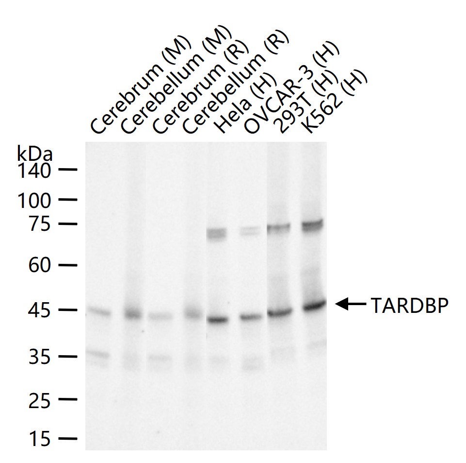
25 ug total protein per lane of various lysates (see on figure) probed with TARDBP monoclonal antibody, unconjugated (bsm-52949R) at 1:2000 dilution and 4°C overnight incubation. Followed by conjugated secondary antibody incubation at r.t. for 60 min.
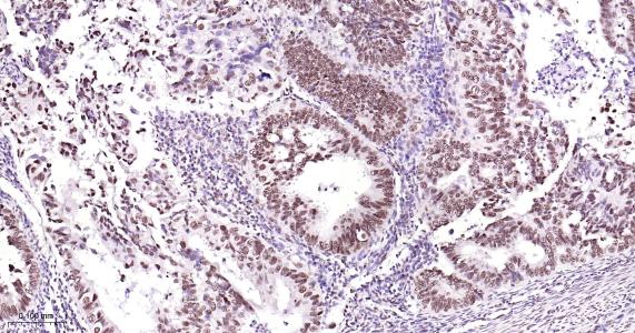
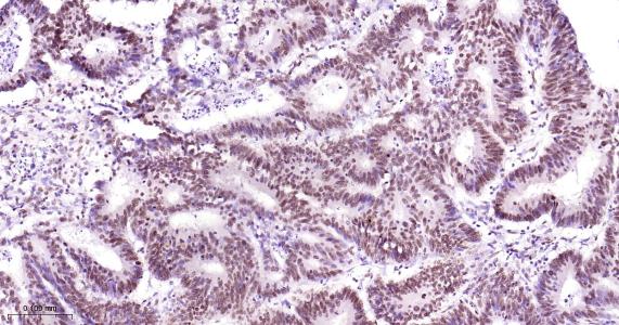
Paraformaldehyde-fixed, paraffin embedded Human Endometrial Cancer; Antigen retrieval by boiling in sodium citrate buffer (pH6.0) for 15 min; Antibody incubation TARDBP Monoclonal Antibody, Unconjugated (bsm-52949R) at 1:200 overnight at 4°C, followed by conjugation to the bs-0295G-HRP and DAB (C-0010) staining.
Paraformaldehyde-fixed, paraffin embedded Human Prostate Cancer; Antigen retrieval by boiling in sodium citrate buffer (pH6.0) for 15 min; Antibody incubation TARDBP Monoclonal Antibody, Unconjugated (bsm-52949R) at 1:200 overnight at 4°C, followed by conjugation to the bs-0295G-HRP and DAB (C-0010) staining.
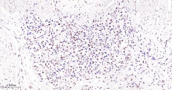
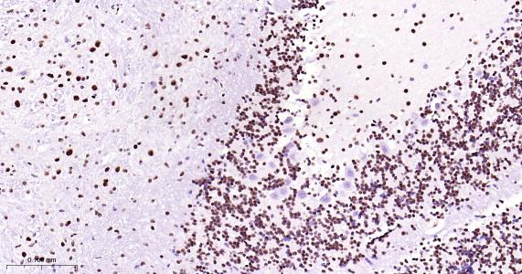
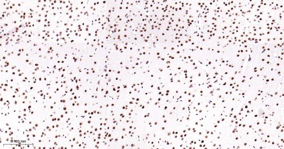
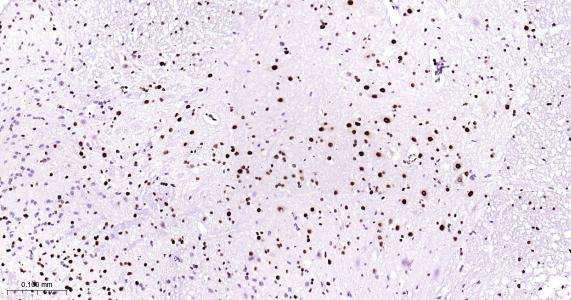
Paraformaldehyde-fixed, paraffin embedded Human Glioma; Antigen retrieval by boiling in sodium citrate buffer (pH6.0) for 15 min; Antibody incubation TARDBP Monoclonal Antibody, Unconjugated (bsm-52949R) at 1:200 overnight at 4°C, followed by conjugation to the bs-0295G-HRP and DAB (C-0010) staining.
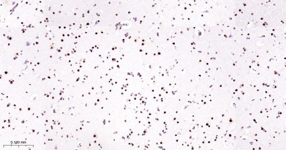
Paraformaldehyde-fixed, paraffin embedded Rat Cerebellum; Antigen retrieval by boiling in sodium citrate buffer (pH6.0) for 15 min; Antibody incubation TARDBP Monoclonal Antibody, Unconjugated (bsm-52949R) at 1:200 overnight at 4°C, followed by conjugation to the bs-0295G-HRP and DAB (C-0010) staining.
Paraformaldehyde-fixed, paraffin embedded Mouse Cerebellum; Antigen retrieval by boiling in sodium citrate buffer (pH6.0) for 15 min; Antibody incubation TARDBP Monoclonal Antibody, Unconjugated (bsm-52949R) at 1:200 overnight at 4°C, followed by conjugation to the bs-0295G-HRP and DAB (C-0010) staining.
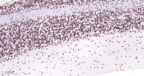
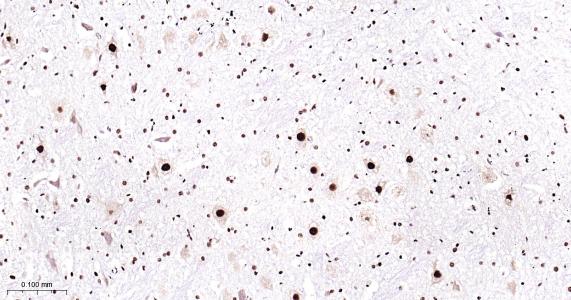
Paraformaldehyde-fixed, paraffin embedded Human Cerebrum; Antigen retrieval by boiling in sodium citrate buffer (pH6.0) for 15 min; Antibody incubation TARDBP Monoclonal Antibody, Unconjugated (bsm-52949R) at 1:200 overnight at 4°C, followed by conjugation to the bs-0295G-HRP and DAB (C-0010) staining.
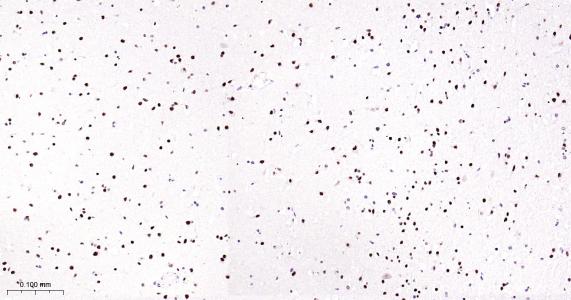
Paraformaldehyde-fixed, paraffin embedded Rat Cerebrum; Antigen retrieval by boiling in sodium citrate buffer (pH6.0) for 15 min; Antibody incubation TARDBP Monoclonal Antibody, Unconjugated (bsm-52949R) at 1:200 overnight at 4°C, followed by conjugation to the bs-0295G-HRP and DAB (C-0010) staining.
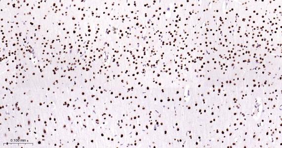
Paraformaldehyde-fixed, paraffin embedded Mouse Cerebrum; Antigen retrieval by boiling in sodium citrate buffer (pH6.0) for 15 min; Antibody incubation TARDBP Monoclonal Antibody, Unconjugated (bsm-52949R) at 1:200 overnight at 4°C, followed by conjugation to the bs-0295G-HRP and DAB (C-0010) staining.
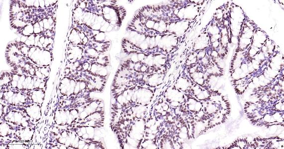
Paraformaldehyde-fixed, paraffin embedded Human Colon Cancer; Antigen retrieval by boiling in sodium citrate buffer (pH6.0) for 15 min; Antibody incubation TARDBP Monoclonal Antibody, Unconjugated (bsm-52949R) at 1:200 overnight at 4°C, followed by conjugation to the bs-0295G-HRP and DAB (C-0010) staining.
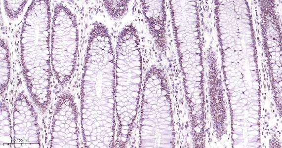
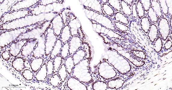
Paraformaldehyde-fixed, paraffin embedded Human Colon; Antigen retrieval by boiling in sodium citrate buffer (pH6.0) for 15 min; Antibody incubation TARDBP Monoclonal Antibody, Unconjugated (bsm-52949R) at 1:200 overnight at 4°C, followed by conjugation to the bs-0295G-HRP and DAB (C-0010) staining.
Paraformaldehyde-fixed, paraffin embedded Rat Colon; Antigen retrieval by boiling in sodium citrate buffer (pH6.0) for 15 min; Antibody incubation TARDBP Monoclonal Antibody, Unconjugated (bsm-52949R) at 1:200 overnight at 4°C, followed by conjugation to the bs-0295G-HRP and DAB (C-0010) staining.
Paraformaldehyde-fixed, paraffin embedded Mouse Colon; Antigen retrieval by boiling in sodium citrate buffer (pH6.0) for 15 min; Antibody incubation TARDBP Monoclonal Antibody, Unconjugated (bsm-52949R) at 1:200 overnight at 4°C, followed by conjugation to the bs-0295G-HRP and DAB (C-0010) staining.
Paraformaldehyde-fixed, paraffin embedded Rat Spinal Cord; Antigen retrieval by boiling in sodium citrate buffer (pH6.0) for 15 min; Antibody incubation TARDBP Monoclonal Antibody, Unconjugated (bsm-52949R) at 1:200 overnight at 4°C, followed by conjugation to the bs-0295G-HRP and DAB (C-0010) staining.
Paraformaldehyde-fixed, paraffin embedded Mouse Spinal Cord; Antigen retrieval by boiling in sodium citrate buffer (pH6.0) for 15 min; Antibody incubation TARDBP Monoclonal Antibody, Unconjugated (bsm-52949R) at 1:200 overnight at 4°C, followed by conjugation to the bs-0295G-HRP and DAB (C-0010) staining.