sales@bioss.com.cn
techsupport@bioss.com.cn
400-901-9800
Host: Rabbit
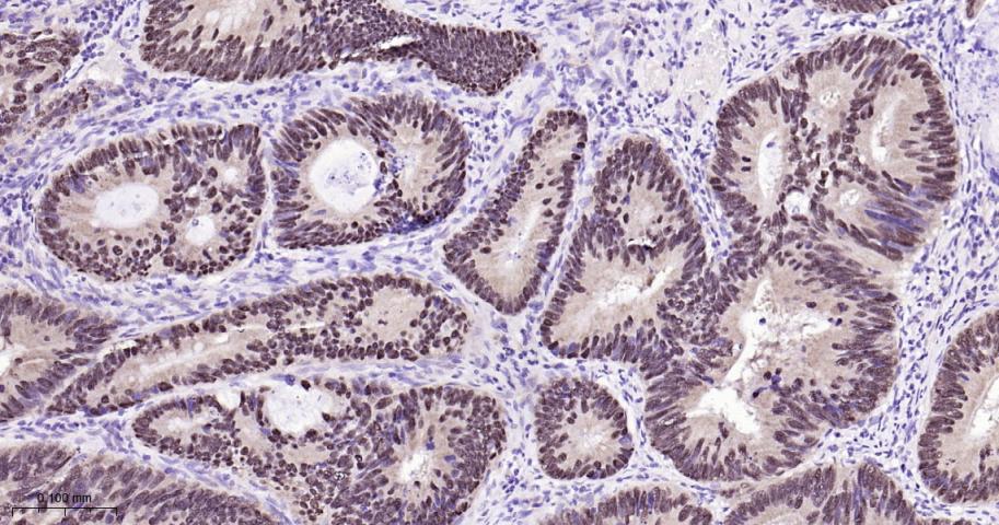
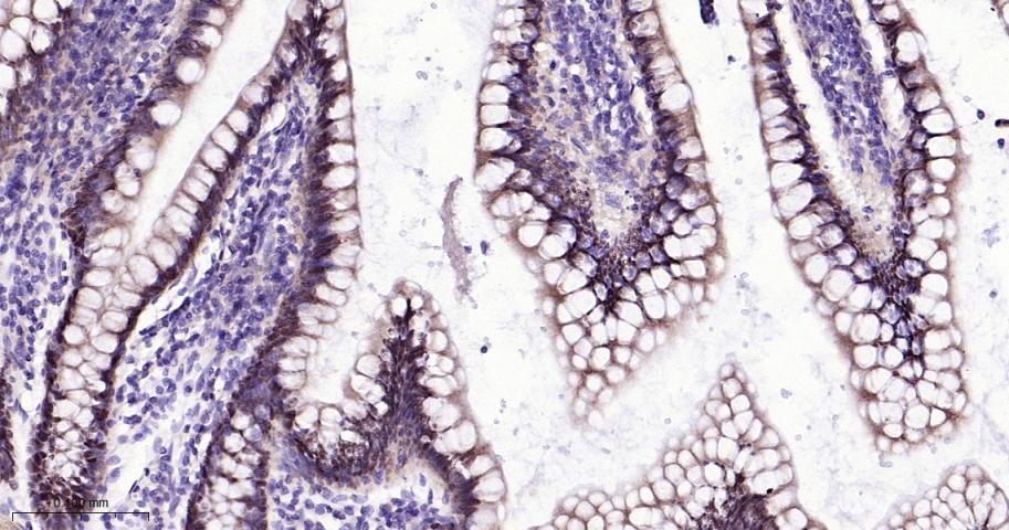
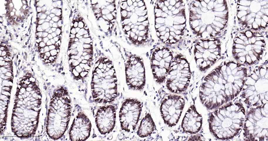
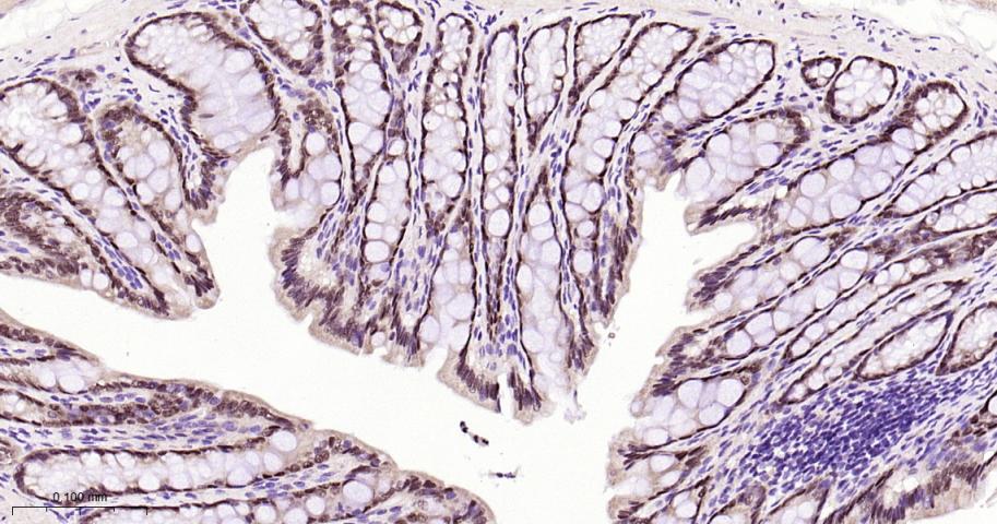
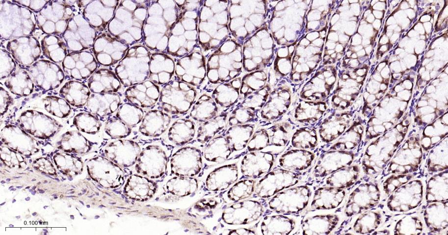
Target Protein: CDX2 Recombinant Rabbit mAb
IR: Immunogen Range:11-76/313
Clonality:
Isotype: IgG
Entrez Gene: 1045
Swiss Prot: Q99626
Source: A synthesized peptide derived from human CDX2:11-76/313
Purification: affinity purified by Protein A
Storage: 0.01M TBS (pH7.4) with 1% BSA, 0.02% Proclin300 and 50% Glycerol. Shipped at 4℃. Store at -20℃ for one year. Avoid repeated freeze/thaw cycles.
Background: This gene is a member of the caudal-related homeobox transcription factor gene family. The encoded protein is a major regulator of intestine-specific genes involved in cell growth an differentiation. This protein also plays a role in early embryonic development of the intestinal tract. Aberrant expression of this gene is associated with intestinal inflammation and tumorigenesis. [provided by RefSeq, Jan 2012]
Size: 100ul
Concentration: 1mg/ml
Applications: IHC-P=1:100-500,IHC-F=1:100-500,IF=1:100-500,Flow-Cyt=1:50-100,ICC/IF=1:50-200
Cross Reactive Species: Human,Mouse,Rat
For research use only. Not intended for diagnostic or therapeutic use.