bsm-52880R [Primary Antibody]
Rabbit  Anti-ADRB2  Antibody
www.bioss.com.cn
sales@bioss.com.cn
techsupport@bioss.com.cn
400-901-9800
DATASHEET

Host: Rabbit

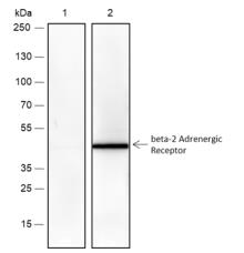
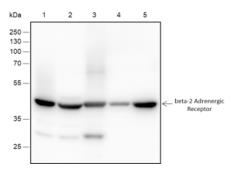
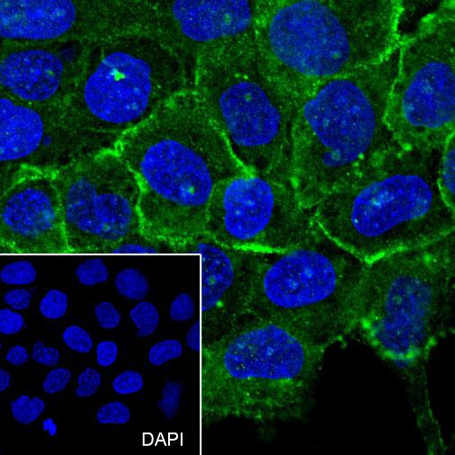
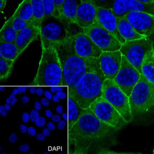
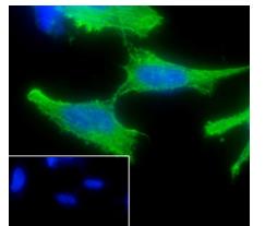
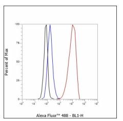
Target Protein: ADRB2

IR: Immunogen Range:

Clonality:

Isotype: IgG

Entrez Gene: 154

Swiss Prot: P07550

Source: KLH conjugated synthetic peptide derived from human ADRB2: 

Purification: affinity purified by Protein A

Storage: 0.01M TBS(pH7.4) with 1% BSA, 0.03% Proclin300 and 50% Glycerol. Shipped at 4℃. Store at -20 °C for one year. Avoid repeated freeze/thaw cycles.

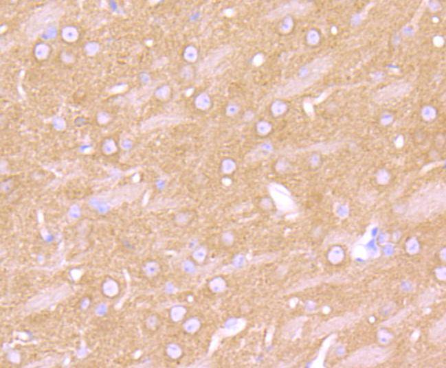
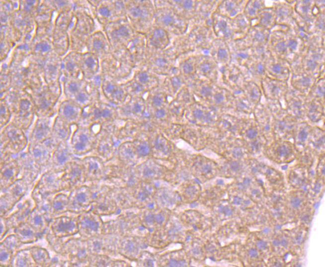
Background: Beta 2 Adrenergic Receptor is a member of the G protein coupled receptor superfamily. This receptor is directly associated with one of its ultimate effectors, the class C L type calcium channel Ca(V)1.2. This receptor channel complex also contains a G protein, an adenylyl cyclase, cAMP dependent kinase, and the counterbalancing phosphatase, PP2A. The assembly of the signaling complex provides a mechanism that ensures specific and rapid signaling by this G protein coupled receptor. This gene contains no introns in either its coding or untranslated sequences. Different polymorphic forms, point mutations, and/or downregulation of this gene are associated with nocturnal asthma, obesity and type 2 diabetes. Expression of the beta 2 Adrenergic Receptor has been reported in adipose, blood, brain, heart, lung, nose, pancreas, skeletal muscle, skin, and vessel.

Size: 100ul

Concentration: 1mg/ml

Applications: WB=1:500-2000,IHC-P=1:100-500,Flow-Cyt=1:100,ICC=1:100

Cross Reactive Species: Human (predicted: Mouse,Rat,Zebrafish)

For research use only. Not intended for diagnostic or therapeutic use.

VALIDATION IMAGES
Western blot analysis of beta 2 Adrenergic Receptor on zebrafish tissue lysates. Proteins were transferred to a PVDF membrane and blocked with 5% BSA in PBS for 1 hour at room temperature. The primary antibody (bsm-52880R, 1/500) was used in 5% BSA at room temperature for 2 hours. Goat Anti-Rabbit IgG - HRP Secondary Antibody at 1:5,000 dilution was used for 1 hour at room temperature.
Blocking buffer: 5% NFDM/TBST Primary Ab dilution: 1:1000 Primary Ab incubation condition: lane 1: PTM-6016+Competitive peptides, lane 2: PTM- 6016, 2 hours at room temperature Secondary Ab: Goat Anti-Rabbit IgG H&L (HRP) Lysate: Mouse kidney Protein loading quantity: 20 μg Exposure time: 60 s Predicted MW: 46 kDa Observed MW: 46 kDa
Blocking buffer: 5% NFDM/TBST Primary Ab dilution: 1:2000 Primary Ab incubation condition: 2 hours at room temperature Secondary Ab: Goat Anti-Rabbit IgG H&L (HRP) Lysate: 1: A431, 2: MCF-7, 3: MDA-MB-231, 4: Rat kidney, 5: Mouse kidney Protein loading quantity: 20 μg Exposure time: 60 s Predicted MW: 46 kDa Observed MW: 46 kDa
Immunohistochemical analysis of paraffin-embedded mouse brain tissue using anti-beta 2 Adrenergic Receptor antibody. The section was pre-treated using heat mediated antigen retrieval with Tris-EDTA buffer (pH 8.0-8.4) for 20 minutes.The tissues were blocked in 5% BSA for 30 minutes at room temperature, washed with ddH2O and PBS, and then probed with the primary antibody (bsm-52880R, 1/50) for 30 minutes at room temperature. The detection was performed using an HRP conjugated compact polymer system. DAB was used as the chromogen. Tissues were counterstained with hematoxylin and mounted with DPX.
Immunohistochemical analysis of paraffin-embedded mouse liver tissue using anti-beta 2 Adrenergic Receptor antibody. The section was pre-treated using heat mediated antigen retrieval with Tris-EDTA buffer (pH 8.0-8.4) for 20 minutes.The tissues were blocked in 5% BSA for 30 minutes at room temperature, washed with ddH2O and PBS, and then probed with the primary antibody (bsm-52880R, 1/50) for 30 minutes at room temperature. The detection was performed using an HRP conjugated compact polymer system. DAB was used as the chromogen. Tissues were counterstained with hematoxylin and mounted with DPX.
Cell line: A431 Fixation: 100% Ice-cold methanol Permeabilization: 0.1% TritonX-100 Primary Ab dilution: 1:100 Primary Ab incubation condition: 4°C overnight Secondary Ab: Goat Anti-Rabbit IgG Nuclear counter stain: DAPI (Blue) Comment: Color green is the positive signal for bsm-52880R
Cell line: MCF7 Fixation: 100% Ice-cold methanol Permeabilization: 0.1% TritonX-100 Primary Ab dilution: 1:100 Primary Ab incubation condition: 4°C overnight Secondary Ab: Goat Anti-Rabbit IgG Nuclear counter stain: DAPI (Blue) Comment: Color green is the positive signal for bsm-52880R
Cell line: SH-SY5Y Fixation: 100% Ice-cold methanol Permeabilization: 0.1% TritonX-100 Primary Ab dilution: 1:50 Primary Ab incubation condition: 4℃ overnight Secondary Ab: Goat Anti-Rabbit IgG Nuclear counter stain: DAPI (Blue) Comment: Color green is the positive signal for PTM-6016
Cell line: SH-SY5Y Fixation: 4% Paraformaldehyde Permeabilization: 90% Methanol Primary Ab dilution: 1:100 Secondary Ab: Goat Anti-Rabbit IgG Unlabelled control: The cell without incubation with primary antibody and secondary antibody (Black line). Isotype control: Rabbit monoclonal IgG (Blue line). Comment: Line red is the positive signal for bsm-52880R
 
PRODUCT SPECIFIC PUBLICATIONS