VALIDATION IMAGES
Blocking buffer: 5% NFDM/TBST
Primary ab dilution: 1:2000
Primary ab incubation condition: 2 hours at room temperature
Secondary ab: Goat Anti-Rabbit IgG H&L (HRP)
Lysate: (-) HeLa, (+) HeLa+Sodium butyrate (30mM, 4hr)
Protein loading quantity: 20 μg
Exposure time: 30 s
Predicted MW: 11 kDa
Observed MW: 11 kDa
Western blot analysis of Histone H4 (acetyl K16) on SiHa cell lysates. Proteins were transferred to a PVDF membrane and blocked with 5% BSA in PBS for 1 hour at room temperature. The primary antibody (bsm-54330R, 1/500) was used in 5% BSA at room temperature for 2 hours. Goat Anti-Rabbit IgG - HRP Secondary Antibody at 1:200,000 dilution was used for 1 hour at room temperature.
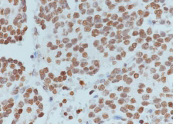
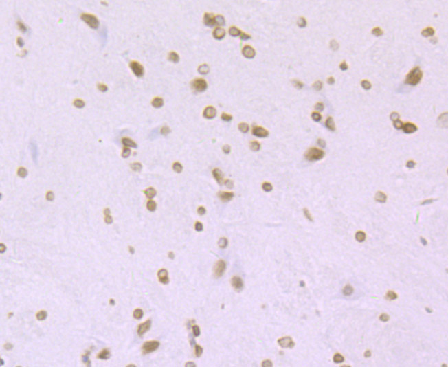
Tissue: Human neuroblastoma
Section type: Formalin fixed & Paraffin-embedded section
Retrieval method: High temperature and high pressure
Retrieval buffer: Tris/EDTA buffer, pH 9.0
Primary ab dilution: 1:200
Primary ab incubation condition: 1 hour at
room temperature
Secondary ab: SP Kit(Rabbit) (sp-0023)
HRP (Ready to use)
Counter stain: Hematoxylin (Blue)
Comment: Color brown is the positive signal for bsm-54330R
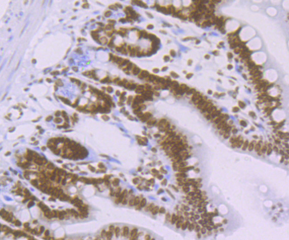
Immunohistochemical analysis of paraffin-embedded mouse colon tissue using anti-Histone H4 (acetyl K16) antibody. The section was pre-treated using heat mediated antigen retrieval with Tris-EDTA buffer (pH 8.0-8.4) for 20 minutes.The tissues were blocked in 5% BSA for 30 minutes at room temperature, washed with ddH2O and PBS, and then probed with the primary antibody (bsm-54330R, 1/50) for 30 minutes at room temperature. The detection was performed using an HRP conjugated compact polymer system. DAB was used as the chromogen. Tissues were counterstained with hematoxylin and mounted with DPX.
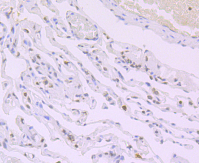
Immunohistochemical analysis of paraffin-embedded human lung carcinoma tissue using anti-Histone H4 (acetyl K16) antibody. The section was pre-treated using heat mediated antigen retrieval with Tris-EDTA buffer (pH 8.0-8.4) for 20 minutes.The tissues were blocked in 5% BSA for 30 minutes at room temperature, washed with ddH2O and PBS, and then probed with the primary antibody (bsm-54330R, 1/50) for 30 minutes at room temperature. The detection was performed using an HRP conjugated compact polymer system. DAB was used as the chromogen. Tissues were counterstained with hematoxylin and mounted with DPX.
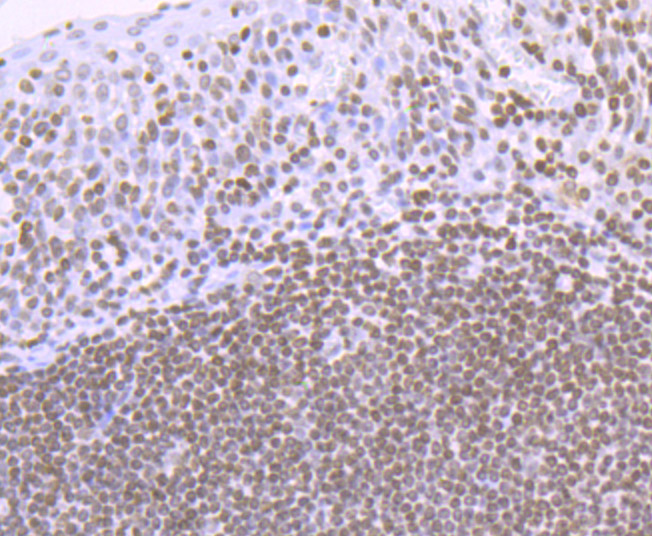
Immunohistochemical analysis of paraffin-embedded human tonsil tissue using anti-Histone H4 (acetyl K16) antibody. The section was pre-treated using heat mediated antigen retrieval with Tris-EDTA buffer (pH 8.0-8.4) for 20 minutes.The tissues were blocked in 5% BSA for 30 minutes at room temperature, washed with ddH2O and PBS, and then probed with the primary antibody (bsm-54330R, 1/50) for 30 minutes at room temperature. The detection was performed using an HRP conjugated compact polymer system. DAB was used as the chromogen. Tissues were counterstained with hematoxylin and mounted with DPX.
Immunohistochemical analysis of paraffin-embedded rat brain tissue using anti-Histone H4 (acetyl K16) antibody. The section was pre-treated using heat mediated antigen retrieval with Tris-EDTA buffer (pH 8.0-8.4) for 20 minutes.The tissues were blocked in 5% BSA for 30 minutes at room temperature, washed with ddH2O and PBS, and then probed with the primary antibody (bsm-54330R, 1/50) for 30 minutes at room temperature. The detection was performed using an HRP conjugated compact polymer system. DAB was used as the chromogen. Tissues were counterstained with hematoxylin and mounted with DPX.
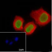
Cell line: HeLa
Fixative: 4% Paraformaldehyde
Permeabilization: 0.1% TritonX-100
Primary ab dilution: 1:200
Primary incubation condition: 4°C overnight
Secondary ab: Goat Anti-Rabbit IgG
Nuclear counter stain: DAPI (Blue)
Counter stain: Tubulin (Red)
Comment: Color green is the positive signal for bsm-54330R
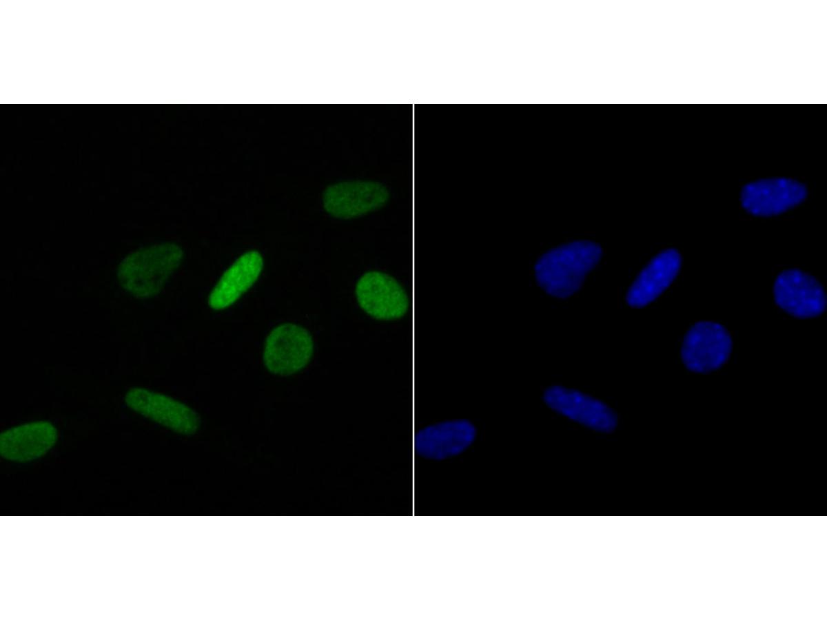
ICC staining of Histone H4 (acetyl K16) in SH-SY5Y cells (green). Formalin fixed cells were permeabilized with 0.1% Triton X-100 in TBS for 10 minutes at room temperature and blocked with 1% Blocker BSA for 15 minutes at room temperature. Cells were probed with the primary antibody (bsm-54330R, 1/50) for 1 hour at room temperature, washed with PBS. Alexa Fluor®488 Goat anti-Rabbit IgG was used as the secondary antibody at 1/1,000 dilution. The nuclear counter stain is DAPI (blue).
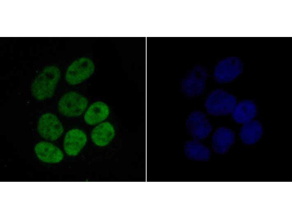
ICC staining of Histone H4 (acetyl K16) in Hela cells (green). Formalin fixed cells were permeabilized with 0.1% Triton X-100 in TBS for 10 minutes at room temperature and blocked with 1% Blocker BSA for 15 minutes at room temperature. Cells were probed with the primary antibody (bsm-54330R, 1/50) for 1 hour at room temperature, washed with PBS. Alexa Fluor®488 Goat anti-Rabbit IgG was used as the secondary antibody at 1/1,000 dilution. The nuclear counter stain is DAPI (blue).