VALIDATION IMAGES
Sample:
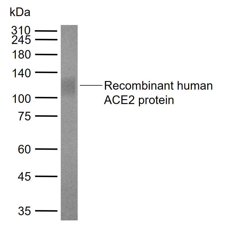
Lane 1: Recombinant human ACE2 protein, His (HEK293)
Primary: Anti-ACE2 (bsm-51221M) at 1/1000 dilution
Secondary: IRDye800CW Goat Anti-Mouse IgG at 1/20000 dilution
Predicted band size: 87 kDa
Observed band size: 110 kDa
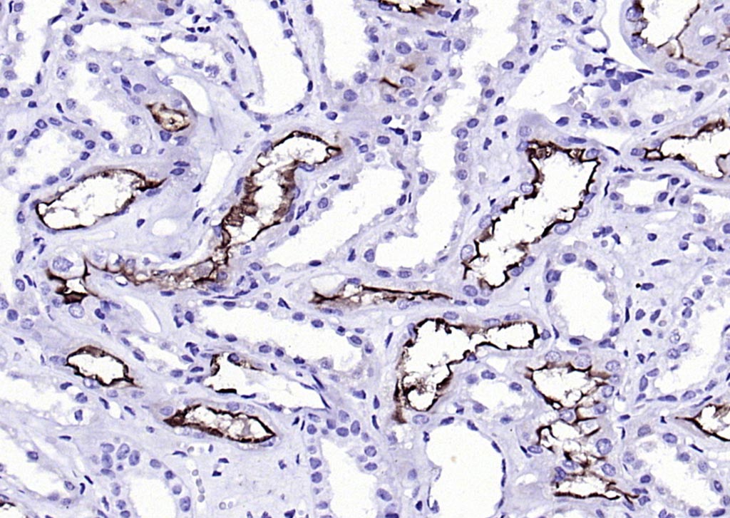
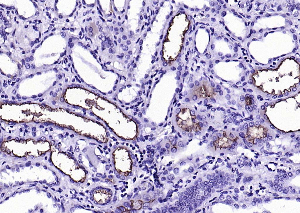
Paraformaldehyde-fixed, paraffin embedded (Human kidney ); Antigen retrieval by boiling in sodium citrate buffer (pH6.0) for 15min; Block endogenous peroxidase by 3% hydrogen peroxide for 20 minutes; Blocking buffer (normal goat serum) at 37°C for 30min; Antibody incubation with (ACE2) Monoclonal Antibody, Unconjugated (bsm-51221M) at 1:200 overnight at 4°C, followed by operating according to SP Kit(Mouse)(sp-0024) instructionsand DAB staining.
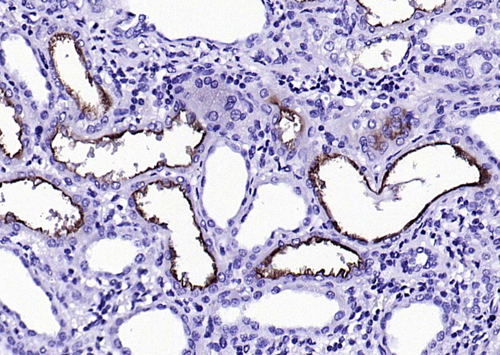
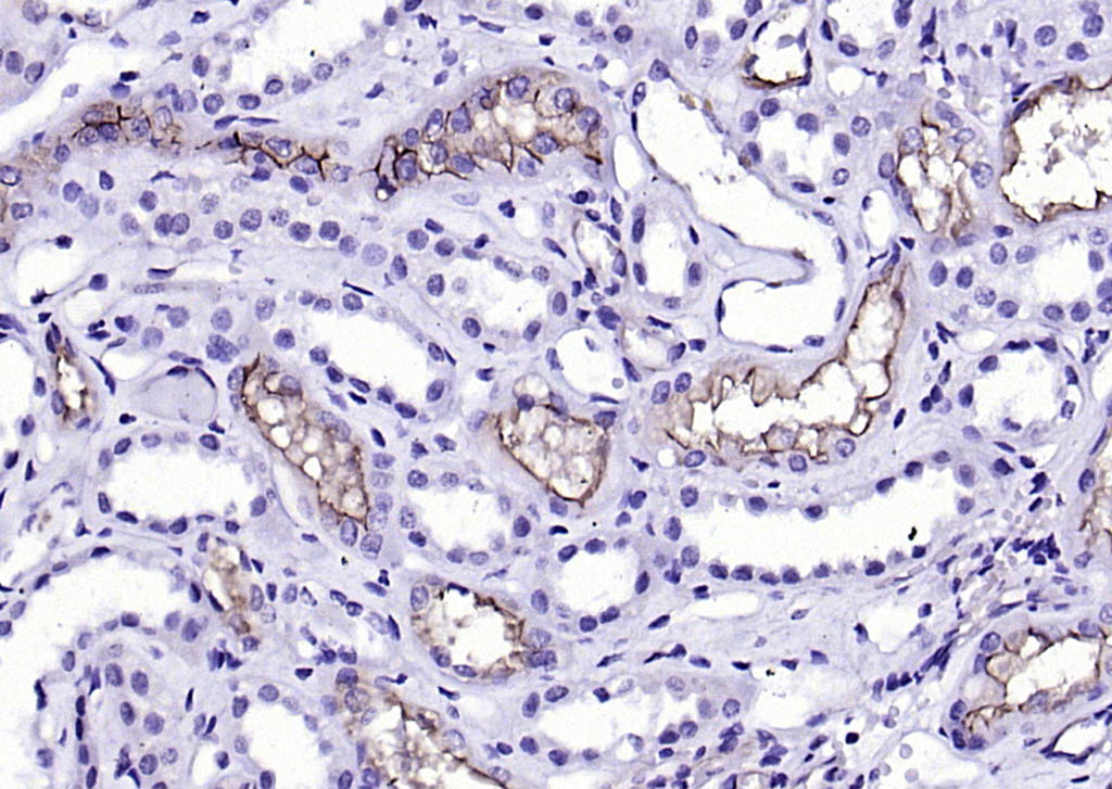
Paraformaldehyde-fixed, paraffin embedded (Human kidney cancer); Antigen retrieval by boiling in sodium citrate buffer (pH6.0) for 15min; Block endogenous peroxidase by 3% hydrogen peroxide for 20 minutes; Blocking buffer (normal goat serum) at 37°C for 30min; Antibody incubation with (ACE2) Monoclonal Antibody, Unconjugated (bsm-51221M) at 1:200 overnight at 4°C, followed by operating according to SP Kit(Mouse)(sp-0024) instructionsand DAB staining.
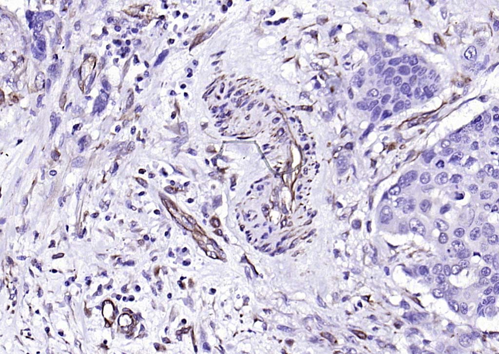
Paraformaldehyde-fixed, paraffin embedded (Human esophageal cancer); Antigen retrieval by boiling in sodium citrate buffer (pH6.0) for 15min; Block endogenous peroxidase by 3% hydrogen peroxide for 20 minutes; Blocking buffer (normal goat serum) at 37°C for 30min; Antibody incubation with (ACE2) Monoclonal Antibody, Unconjugated (bsm-51221M) at 1:200 overnight at 4°C, followed by operating according to SP Kit(Mouse)(sp-0024) instructionsand DAB staining.
Paraformaldehyde-fixed, paraffin embedded (Human kidney cancer); Antigen retrieval by boiling in sodium citrate buffer (pH6.0) for 15min; Block endogenous peroxidase by 3% hydrogen peroxide for 20 minutes; Blocking buffer (normal goat serum) at 37°C for 30min; Antibody incubation with (ACE2) Monoclonal Antibody, Unconjugated (bsm-51221M) at 1:200 overnight at 4°C, followed by operating according to SP Kit(Mouse)(sp-0024) instructionsand DAB staining.
Paraformaldehyde-fixed, paraffin embedded (Human kidney ); Antigen retrieval by boiling in sodium citrate buffer (pH6.0) for 15min; Block endogenous peroxidase by 3% hydrogen peroxide for 20 minutes; Blocking buffer (normal goat serum) at 37°C for 30min; Antibody incubation with (ACE2) Monoclonal Antibody, Unconjugated (bsm-51221M) at 1:200 overnight at 4°C, followed by operating according to SP Kit(Mouse)(sp-0024) instructionsand DAB staining.
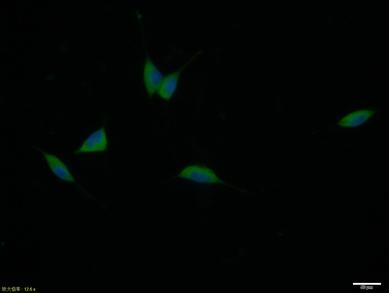
SH-SY5Y cell; 4% Paraformaldehyde-fixed; Triton X-100 at room temperature for 20 min; Blocking buffer (normal goat serum, C-0005) at 37°C for 20 min; Antibody incubation with (ACE2) monoclonal Antibody, Unconjugated (bsm-51221M) 1:100, 90 minutes at 37°C; followed by a conjugated Goat Anti-Mouse IgG antibody at 37°C for 90 minutes, DAPI (blue, C02-04002) was used to stain the cell nuclei.