bsm-52811R [Primary Antibody]
Rabbit  Anti-LYVE1  Antibody
www.bioss.com.cn
sales@bioss.com.cn
techsupport@bioss.com.cn
400-901-9800
DATASHEET

Host: Rabbit

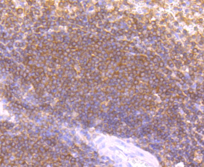
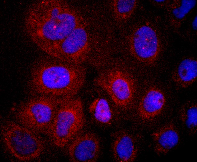
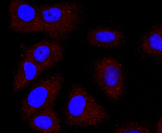
Target Protein: LYVE1

IR: Immunogen Range:

Clonality:

Isotype: IgG

Entrez Gene: 10894

Swiss Prot: Q9Y5Y7

Source: Recombinant human LYVE1 protein: 

Purification: affinity purified by Protein A

Storage: 0.01M TBS(pH7.4) with 1% BSA, 0.03% Proclin300 and 50% Glycerol. Shipped at 4℃. Store at -20 °C for one year. Avoid repeated freeze/thaw cycles.

Background: The lymphatic vasculature forms a second circulatory system that drains extracellular fluid from the tissues and provides an exclusive environment in which immune cells can encounter and respond to foreign antigen. Recently a number of interesting molecules have been identified that may be exploited as markers for lymphatic endothelium, including the hyaluronan receptor LYVE1, PALE, VEGFR3, podoplanin. LYVE1 has been identified as a major receptor for HA (extracellular matrix glycosaminoglycan hyaluronan) on the lymph vessel wall. The deduced amino acid sequence of LYVE1 predicts a 322-residue type I integral membrane polypeptide 41% similar to the CD44 HA receptor with a 212-residue extracellular domain containing a single Link module the prototypic HA binding domain of the Link protein superfamily. Like CD44, the LYVE1 molecule binds both soluble and immobilized HA. However, unlike CD44, the LYVE1 molecule colocalizes with HA on the luminal face of the lymph vessel wall and is completely absent from blood vessels. Hence, LYVE1 is the first lymph-specific HA receptor to be characterized and is a uniquely powerful marker for lymph vessels themselves.

Size: 100ul

Concentration: 1mg/ml

Applications: IHC-P=1:50-200,IHC-F=1:50-200,Flow-Cyt=1:50,ICC=1:50

Cross Reactive Species: Human

For research use only. Not intended for diagnostic or therapeutic use.

VALIDATION IMAGES
Sample: Lane 1: Mouse Spleen tissue lysates Lane 2: Mouse Blood cell lysates Lane 3: Mouse Lymph node tissue lysates Lane 4: Mouse Large intestine tissue lysates Lane 5: Rat Thyroid gland tissue lysates Lane 6: Rat Spleen tissue lysates Lane 7: Rat Lymph node tissue lysates Lane 8: Rat Large intestine tissue lysates Lane 9: Human Huvec cell lysates Lane 10: Human K562 cell lysates Primary: Anti- LYVE-1 (bsm-52811R) at 1/1000 dilution Secondary: IRDye800CW Goat Anti-Rabbit IgG at 1/20000 dilution Predicted band size: 32 kD Observed band size: 58 kD
Paraformaldehyde-fixed, paraffin embedded (human spleen tissue); Antigen retrieval by boiling in sodium citrate buffer (pH6.0) for 15min; Block endogenous peroxidase by 3% hydrogen peroxide for 20 minutes; Blocking buffer (normal goat serum) at 37°C for 30min; Antibody incubation with (LYVE1) Monoclonal Antibody, Unconjugated (bsm-52811R) at 1:50 overnight at 4°C, followed by operating according to SP Kit(Rabbit) (sp-0023) instructionsand DAB staining.
A431 cell; 4% Paraformaldehyde-fixed; Triton X-100 at room temperature for 20 min; Blocking buffer (normal goat serum,C-0005) at 37°C for 20 min; Antibody incubation with (LYVE1) monoclonal Antibody, Unconjugated (bsm-52811R) 1:50, 90 minutes at 37°C; followed by a conjugated Goat Anti-Rabbit IgG antibody at 37°C for 90 minutes, DAPI (blue, C02-04002) was used to stain the cell nuclei.
SW480 cell; 4% Paraformaldehyde-fixed; Triton X-100 at room temperature for 20 min; Blocking buffer (normal goat serum,C-0005) at 37°C for 20 min; Antibody incubation with (LYVE1) monoclonal Antibody, Unconjugated (bsm-52811R) 1:50, 90 minutes at 37°C; followed by a conjugated Goat Anti-Rabbit IgG antibody at 37°C for 90 minutes, DAPI (blue, C02-04002) was used to stain the cell nuclei.
HUVEC cell; 4% Paraformaldehyde-fixed; Triton X-100 at room temperature for 20 min; Blocking buffer (normal goat serum,C-0005) at 37°C for 20 min; Antibody incubation with (LYVE1) monoclonal Antibody, Unconjugated (bsm-52811R) 1:50, 90 minutes at 37°C; followed by a conjugated Goat Anti-Rabbit IgG antibody at 37°C for 90 minutes, DAPI (blue, C02-04002) was used to stain the cell nuclei.
Blank control:Hela. Primary Antibody (green line): Rabbit Anti-LYVE1 antibody (bsm-52811R) Dilution: 1:50; Isotype Control Antibody (orange line): Rabbit IgG . Secondary Antibody : Goat anti-rabbit IgG-AF488 Dilution: 1:1000. Protocol The cells were fixed with 4% PFA (10min at room temperature)and then permeabilized with 0.1% PBST for 20 min at room temperature.The cells were then incubated in 5%BSA to block non-specific protein-protein interactions for 30 min at room temperature .Cells stained with Primary Antibody for 30 min at room temperature. The secondary antibody used for 40 min at room temperature. Acquisition of 20,000 events was performed.
 
PRODUCT SPECIFIC PUBLICATIONS