sales@bioss.com.cn
techsupport@bioss.com.cn
400-901-9800
Host: Rabbit
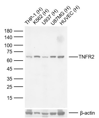
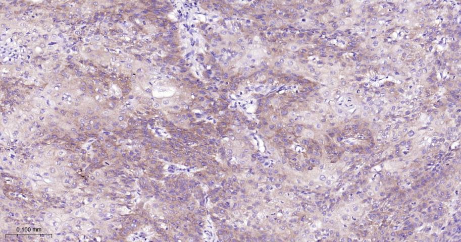
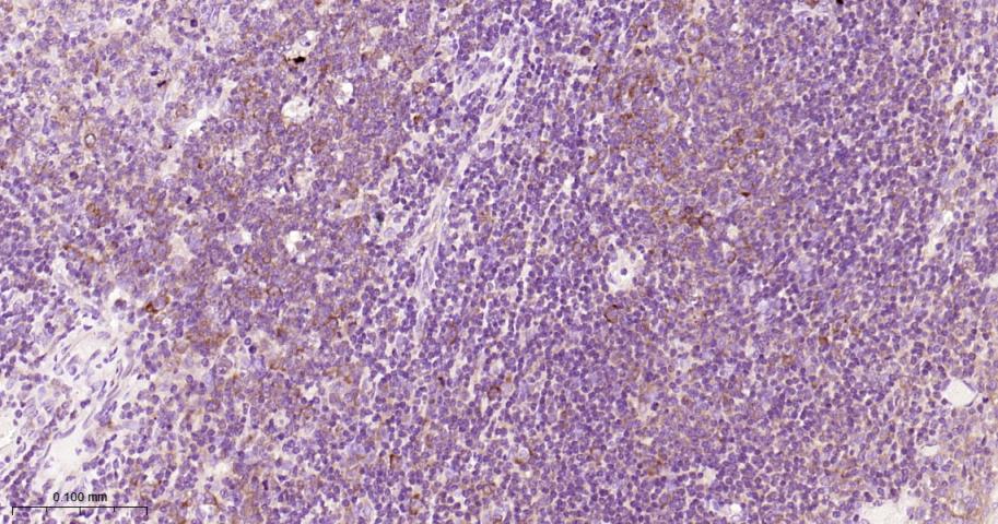
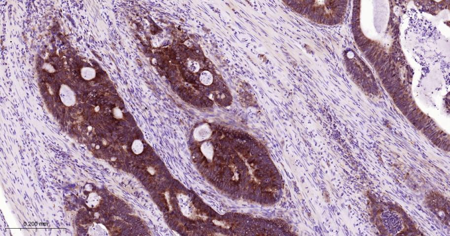
Target Protein: TNFR2 Recombinant Rabbit mAb
IR: Immunogen Range:415-461/461
Clonality:
Isotype: IgG
Entrez Gene: 7133
Swiss Prot: P20333
Source: A synthesized peptide derived from human TNFRSF1B:415-461/461
Purification: affinity purified by Protein A
Storage: 0.01M TBS (pH7.4) with 1% BSA, 0.02% Proclin300 and 50% Glycerol. Shipped at 4℃. Store at -20℃ for one year. Avoid repeated freeze/thaw cycles.
Background: The protein encoded by this gene is a member of the TNF-receptor superfamily. This protein and TNF-receptor 1 form a heterocomplex that mediates the recruitment of two anti-apoptotic proteins, c-IAP1 and c-IAP2, which possess E3 ubiquitin ligase activity. The function of IAPs in TNF-receptor signalling is unknown, however, c-IAP1 is thought to potentiate TNF-induced apoptosis by the ubiquitination and degradation of TNF-receptor-associated factor 2, which mediates anti-apoptotic signals. Knockout studies in mice also suggest a role of this protein in protecting neurons from apoptosis by stimulating antioxidative pathways. [provided by RefSeq, Jul 2008]
Size: 100ul
Concentration: 1mg/ml
Applications: IHC-P=1:50-200,IHC-F=1:50-200,IF=1:50-200
Cross Reactive Species: Human (predicted: Mouse,Rat)
For research use only. Not intended for diagnostic or therapeutic use.