VALIDATION IMAGES
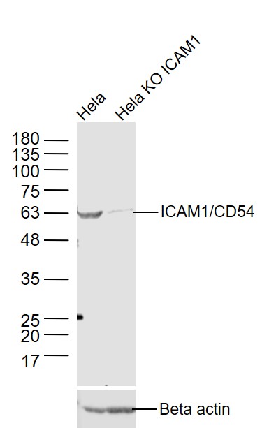
Sample:
Hela(Human) Cell Lysate at 30 ug
Hela KO ICAM1 (Human) Cell Lysate at 30 ug
Primary: Anti-ICAM1/CD54 (bs-0608R) at 1/1000 dilution
Secondary: IRDye800CW Goat Anti-Rabbit IgG at 1/20000 dilution
Predicted band size: 56 kD
Observed band size: 56 kD
Sample:
A549(Human) Cell Lysate at 30 ug
Primary: Anti-ICAM1 (bs-0608R) at 1/300 dilution
Secondary: IRDye800CW Goat Anti-Rabbit IgG at 1/20000 dilution
Predicted band size: 56 kD
Observed band size: 56/69 kD
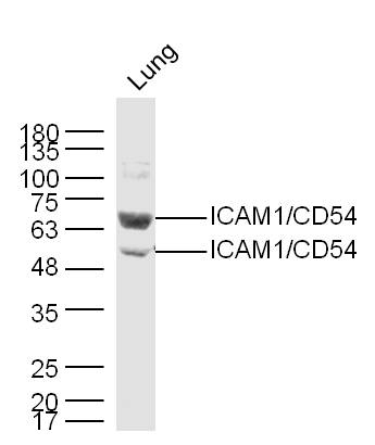
Sample:
Lung (Mouse) Lysate at 40 ug
Primary: Anti-ICAM1 (bs-0608R) at 1/300 dilution
Secondary: IRDye800CW Goat Anti-Rabbit IgG at 1/20000 dilution
Predicted band size: 56 kD
Observed band size: 56/69 kD
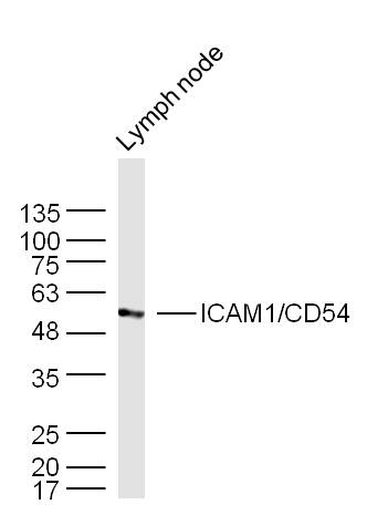
Sample:
Lymph node (Mouse) Lysate at 40 ug
Primary: Anti-ICAM1 (bs-0608R) at 1/300 dilution
Secondary: IRDye800CW Goat Anti-Rabbit IgG at 1/20000 dilution
Predicted band size: 56 kD
Observed band size: 56 kD
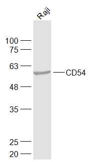
Sample:
Raji(Human) Cell Lysate at 30 ug
Primary: Anti-CD54 (bs-0608R) at 1/1000 dilution
Secondary: IRDye800CW Goat Anti-Rabbit IgG at 1/20000 dilution
Predicted band size: 56 kD
Observed band size: 56 kD
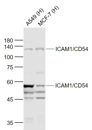
Sample:
Lane 1: A549 (Human) Cell Lysate at 30 ug
Lane 2: MCF-7 (Human) Cell Lysate at 30 ug
Primary: Anti-ICAM1/CD54 (bs-0608R) at 1/1000 dilution
Secondary: IRDye800CW Goat Anti-Rabbit IgG at 1/20000 dilution
Predicted band size: 110/58 kD
Observed band size: 110/58 kD
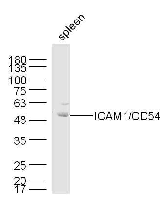
Sample:
Spleen (Mouse) Lysate at 40 ug
Primary: Anti-ICAM1 (bs-0608R) at 1/300 dilution
Secondary: IRDye800CW Goat Anti-Rabbit IgG at 1/20000 dilution
Predicted band size: 56 kD
Observed band size: 56/69 kD
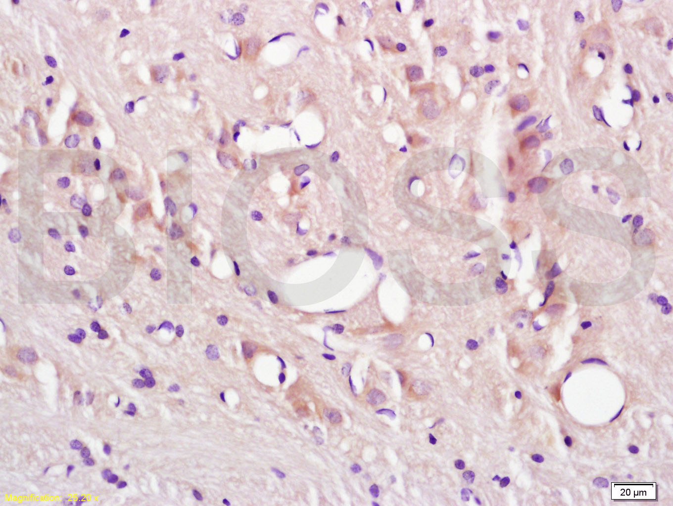
Tissue/cell: rat brain tissue; 4% Paraformaldehyde-fixed and paraffin-embedded;
Antigen retrieval: citrate buffer ( 0.01M, pH 6.0 ), Boiling bathing for 15min; Block endogenous peroxidase by 3% Hydrogen peroxide for 30min; Blocking buffer (normal goat serum,C-0005) at 37℃ for 20 min;
Incubation: Anti-CD54/ICAM-1 Polyclonal Antibody, Unconjugated(bs-0608R) 1:200, overnight at 4°C, followed by conjugation to the secondary antibody(SP-0023) and DAB(C-0010) staining
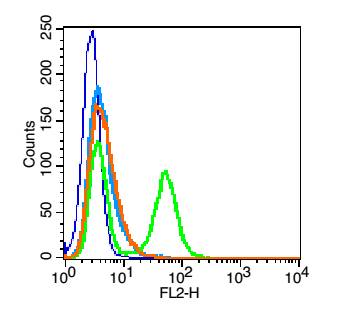
Blank control (blue line): A431 cells(blue).
Primary Antibody (green line): Rabbit Anti-ICAM1/PE-CY7 Conjugated antibody (bs-0608R-PE-CY7)
Dilution: 1μg /10^6 cells;
Isotype Control Antibody (orange line): Rabbit IgG-PE-CY7 .
Protocol
The cells were fixed with 70% ice-cold methanol overnight at 4℃ . The cells were then incubated in 1 X PBS/2%BSA/10% goat serum to block non-specific protein-protein interactions followed by the antibody for 15 min at room temperature. Cells stained with Primary Antibody for 30 min at room temperature.Acquisition of 20,000 events was performed.
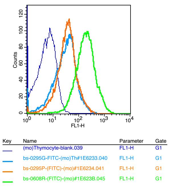
Blank control: mouse thymouses(blue)
Isotype Control Antibody: Rabbit IgG(orange) ;
Secondary Antibody: Goat anti-rabbit IgG-FITC(white blue),
Dilution: 1:100 in 1 X PBS containing 0.5% BSA ;
Primary Antibody Dilution: 1μg in 100 μL 1X PBS containing 0.5% BSA(green).
Blank control: HUVEC cells(blue). Primary Antibody:Rabbit Anti-CD54 antibody(bs-0608R), Dilution: 1μg in 100 μL 1X PBS containing 0.5% BSA; Isotype Control Antibody: Rabbit IgG(orange) ,used under the same conditions ); Secondary Antibody: Goat anti-rabbit IgG-PE(white blue), Dilution: 1:200 in 1 X PBS containing 0.5% BSA.
Protocol
The cells were fixed with 2% paraformaldehyde (10 min) .Primary antibody (bs-0608R, 1μg /1x10^6 cells) were incubated for 30 min on the ice, followed by 1 X PBS containing 0.5% BSA + 1 0% goat serum (15 min) to block non-specific protein-protein interactions. Then the Goat Anti-rabbit IgG/PE antibody was added into the blocking buffer mentioned above to react with the primary antibody at 1/200 dilution for 30 min on ice. Acquisition of 20,000 events was performed.