VALIDATION IMAGES
Sample:
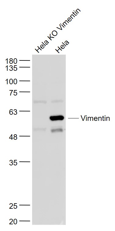
Hela KO Vimentin (Human) Cell Lysate at 30 ug
Hela(Human) Cell Lysate at 30 ug
Primary: Anti- Vimentin (bs-8533R) at 1/1000 dilution
Secondary: IRDye800CW Goat Anti-Rabbit IgG at 1/20000 dilution
Predicted band size: 51 kD
Observed band size: 57 kD
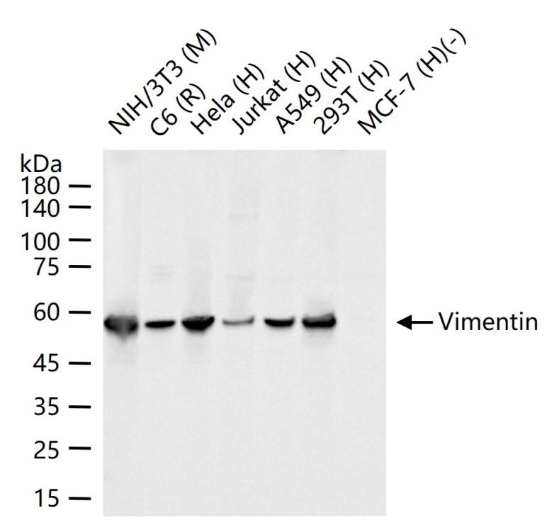
25 ug total protein per lane of various lysates (see on figure) probed with Vimentin polyclonal antibody, unconjugated (bs-8533R) at 1:1000 dilution and 4°C overnight incubation. Followed by conjugated secondary antibody incubation at r.t. for 60 min.
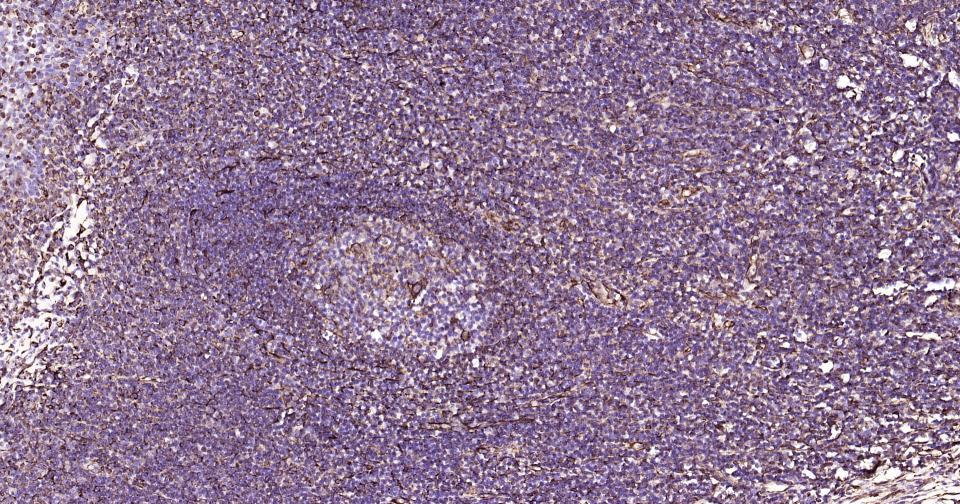
Paraformaldehyde-fixed, paraffin embedded Human Tonsil; Antigen retrieval by boiling in sodium citrate buffer (pH6.0) for 15 min; Antibody incubation with Vimentin Polyclonal Antibody, Unconjugated (bs-8533R) at 1:200 overnight at 4°C, followed by conjugation to the SP Kit (Rabbit, SP-0023) and DAB (C-0010) staining.
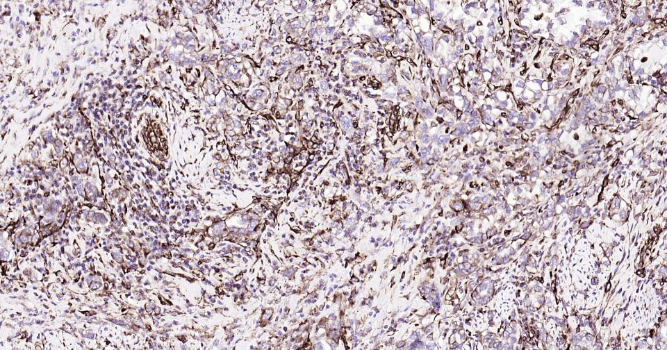
Paraformaldehyde-fixed, paraffin embedded Human Cervical Cancer; Antigen retrieval by boiling in sodium citrate buffer (pH6.0) for 15 min; Antibody incubation with Vimentin Polyclonal Antibody, Unconjugated (bs-8533R) at 1:200 overnight at 4°C, followed by conjugation to the SP Kit (Rabbit, SP-0023) and DAB (C-0010) staining.
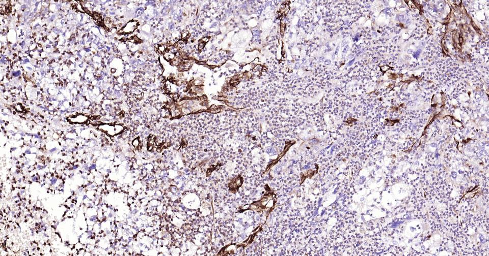
Paraformaldehyde-fixed, paraffin embedded Human Lung Cancer; Antigen retrieval by boiling in sodium citrate buffer (pH6.0) for 15 min; Antibody incubation with Vimentin Polyclonal Antibody, Unconjugated (bs-8533R) at 1:200 overnight at 4°C, followed by conjugation to the SP Kit (Rabbit, SP-0023) and DAB (C-0010) staining.
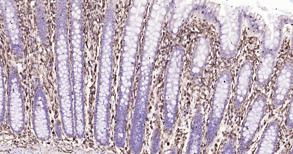
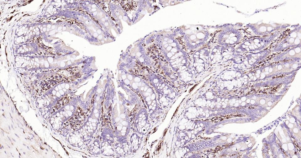
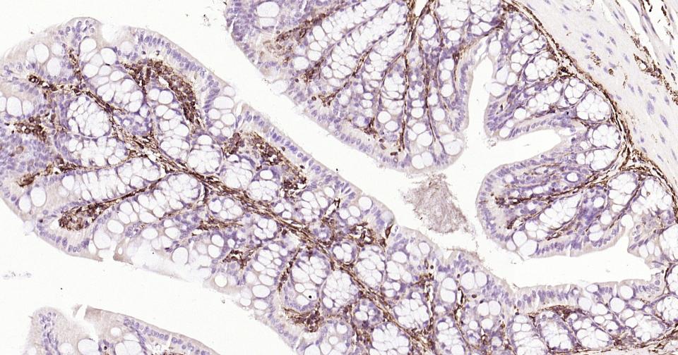
Paraformaldehyde-fixed, paraffin embedded Human Colon; Antigen retrieval by boiling in sodium citrate buffer (pH6.0) for 15 min; Antibody incubation with Vimentin Polyclonal Antibody, Unconjugated (bs-8533R) at 1:200 overnight at 4°C, followed by conjugation to the SP Kit (Rabbit, SP-0023) and DAB (C-0010) staining.
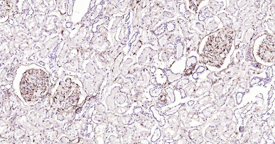
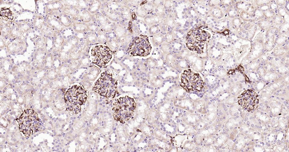
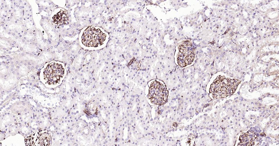
Paraformaldehyde-fixed, paraffin embedded Human Kidney; Antigen retrieval by boiling in sodium citrate buffer (pH6.0) for 15 min; Antibody incubation with Vimentin Polyclonal Antibody, Unconjugated (bs-8533R) at 1:200 overnight at 4°C, followed by conjugation to the SP Kit (Rabbit, SP-0023) and DAB (C-0010) staining.
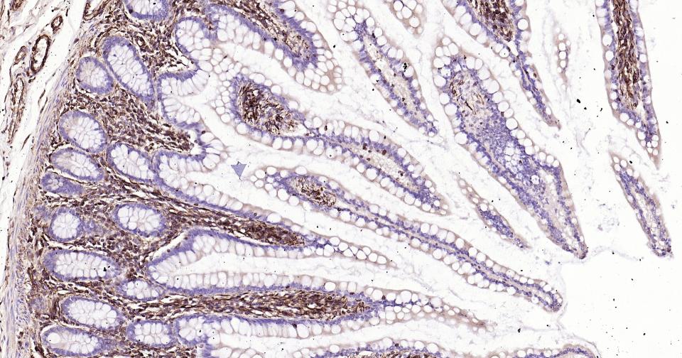
Paraformaldehyde-fixed, paraffin embedded Human Small Intestine; Antigen retrieval by boiling in sodium citrate buffer (pH6.0) for 15 min; Antibody incubation with Vimentin Polyclonal Antibody, Unconjugated (bs-8533R) at 1:200 overnight at 4°C, followed by conjugation to the SP Kit (Rabbit, SP-0023) and DAB (C-0010) staining.
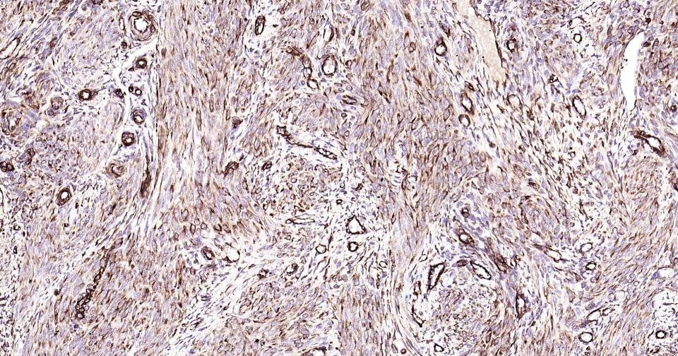
Paraformaldehyde-fixed, paraffin embedded Human Uterus; Antigen retrieval by boiling in sodium citrate buffer (pH6.0) for 15 min; Antibody incubation with Vimentin Polyclonal Antibody, Unconjugated (bs-8533R) at 1:200 overnight at 4°C, followed by conjugation to the SP Kit (Rabbit, SP-0023) and DAB (C-0010) staining.
Paraformaldehyde-fixed, paraffin embedded Human Breast Cancer; Antigen retrieval by boiling in sodium citrate buffer (pH6.0) for 15 min; Antibody incubation with Vimentin Polyclonal Antibody, Unconjugated (bs-8533R) at 1:200 overnight at 4°C, followed by conjugation to the SP Kit (Rabbit, SP-0023) and DAB (C-0010) staining.
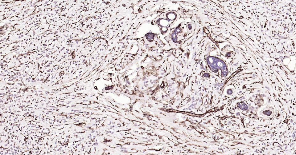
Paraformaldehyde-fixed, paraffin embedded Human Endometrium Cancer; Antigen retrieval by boiling in sodium citrate buffer (pH6.0) for 15 min; Antibody incubation with Vimentin Polyclonal Antibody, Unconjugated (bs-8533R) at 1:200 overnight at 4°C, followed by conjugation to the SP Kit (Rabbit, SP-0023) and DAB (C-0010) staining.
Paraformaldehyde-fixed, paraffin embedded Mouse Kidney; Antigen retrieval by boiling in sodium citrate buffer (pH6.0) for 15 min; Antibody incubation with Vimentin Polyclonal Antibody, Unconjugated (bs-8533R) at 1:200 overnight at 4°C, followed by conjugation to the SP Kit (Rabbit, SP-0023) and DAB (C-0010) staining.
Paraformaldehyde-fixed, paraffin embedded Rat Colon; Antigen retrieval by boiling in sodium citrate buffer (pH6.0) for 15 min; Antibody incubation with Vimentin Polyclonal Antibody, Unconjugated (bs-8533R) at 1:200 overnight at 4°C, followed by conjugation to the SP Kit (Rabbit, SP-0023) and DAB (C-0010) staining.
Paraformaldehyde-fixed, paraffin embedded Mouse Colon; Antigen retrieval by boiling in sodium citrate buffer (pH6.0) for 15 min; Antibody incubation with Vimentin Polyclonal Antibody, Unconjugated (bs-8533R) at 1:200 overnight at 4°C, followed by conjugation to the SP Kit (Rabbit, SP-0023) and DAB (C-0010) staining.
Paraformaldehyde-fixed, paraffin embedded Rat Kidney; Antigen retrieval by boiling in sodium citrate buffer (pH6.0) for 15 min; Antibody incubation with Vimentin Polyclonal Antibody, Unconjugated (bs-8533R) at 1:200 overnight at 4°C, followed by conjugation to the SP Kit (Rabbit, SP-0023) and DAB (C-0010) staining.
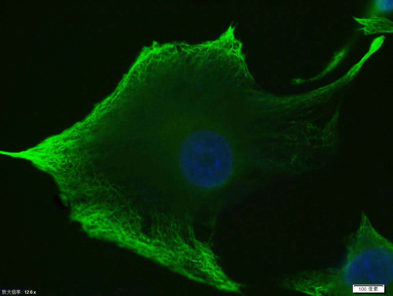
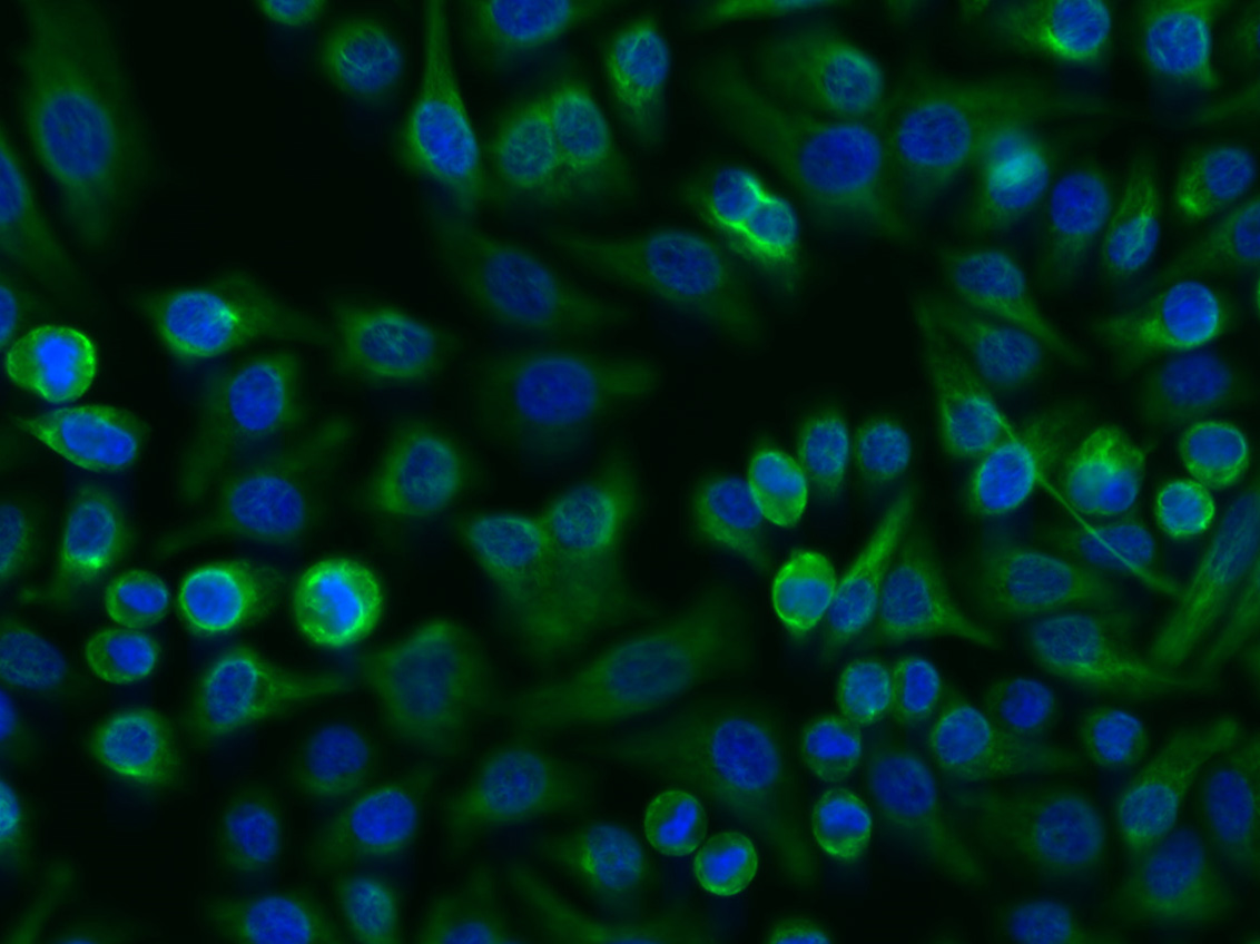
Tissue/cell: U-87MG cell; 4% Paraformaldehyde-fixed; Triton X-100 at room temperature for 20 min; Blocking buffer (normal goat serum, C-0005) at 37°C for 20 min; Antibody incubation with (Vimentin) Polyclonal Antibody, Unconjugated (bs-8533R)antibody (bs-0295G-FITC) at 37°C for 90 minutes, DAPI (blue, C02-04002) was used to stain the cell nuclei.
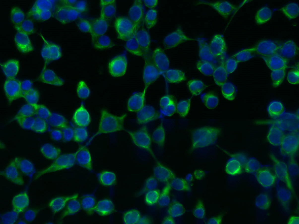
Tissue/cell: 293T cell; 4% Paraformaldehyde-fixed; Triton X-100 at room temperature for 20 min; Blocking buffer (normal goat serum, C-0005) at 37°C for 20 min; Antibody incubation with (Vimentin) Polyclonal Antibody, Unconjugated (bs-8533R) 1:200, 2 hours at 37°C; followed by a conjugated Goat Anti-Rabbit IgG antibody (bs-0295G-FITC) at 37°C for 90 minutes, DAPI (5ug/ml, blue, C-0033) was used to stain the cell nuclei.
Tissue/cell: FHC cell; 4% Paraformaldehyde-fixed; Triton X-100 at room temperature for 20 min; Blocking buffer (normal goat serum, C-0005) at 37°C for 20 min; Antibody incubation with (Vimentin) Polyclonal Antibody, Unconjugated (bs-8533R) 1:200, 2 hours at 37°C; followed by a conjugated Goat Anti-Rabbit IgG antibody (bs-0295G-FITC) at 37°C for 90 minutes, DAPI (5ug/ml, blue, C-0033) was used to stain the cell nuclei.
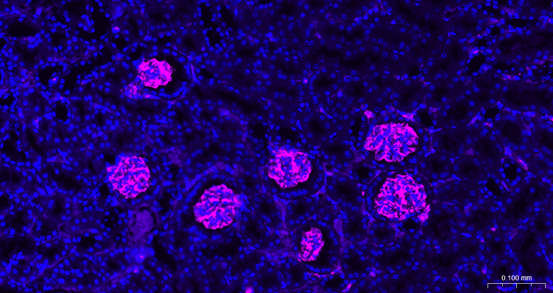
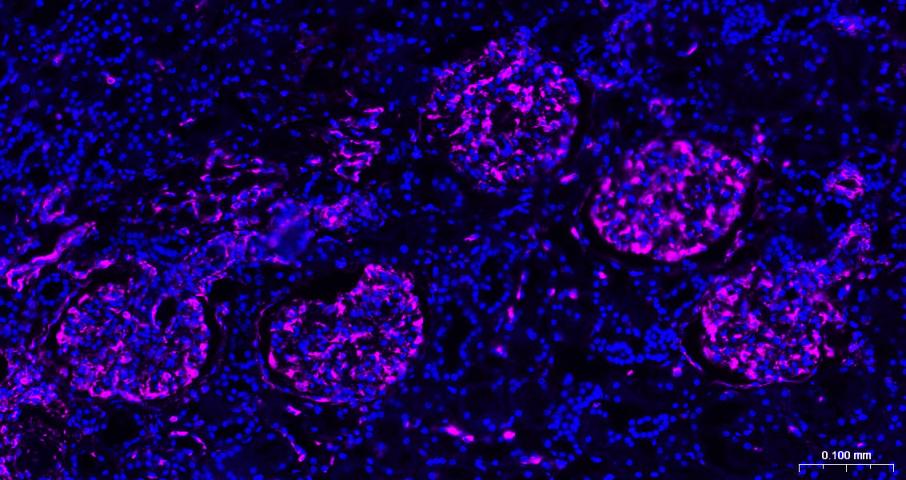
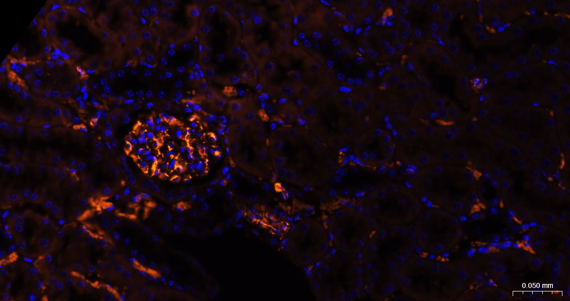
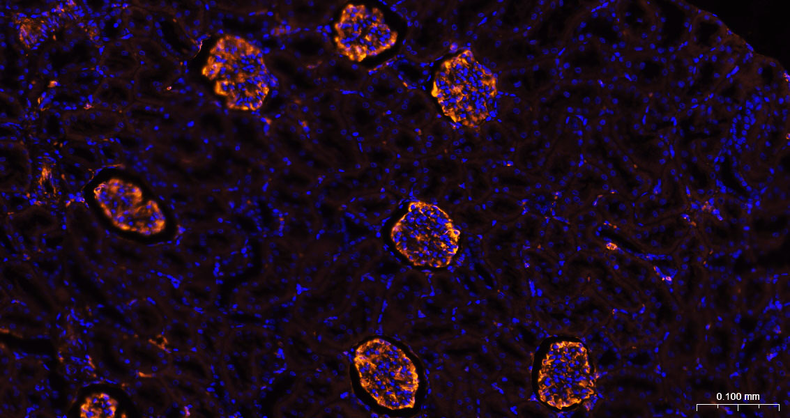
Paraformaldehyde-fixed, paraffin embedded Mouse Kidney; Antigen retrieval by boiling in sodium citrate buffer (pH6.0) for 15 min; The section was incubated with Vimentin Polyclonal Antibody, Unconjugated (bs-8533R) at 1:200 overnight at 4°C. Followed by conjugated Goat Anti-Rabbit IgG antibody (Rose Red, bs-40295G-BF647), DAPI (blue, C02-04002) was used to stain the cell nuclei.
Paraformaldehyde-fixed, paraffin embedded Rat Kidney; Antigen retrieval by boiling in sodium citrate buffer (pH6.0) for 15 min; The section was incubated with Vimentin Polyclonal Antibody, Unconjugated (bs-8533R) at 1:200 overnight at 4°C. Followed by conjugated Goat Anti-Rabbit IgG antibody (Rose Red, bs-40295G-BF647), DAPI (blue, C02-04002) was used to stain the cell nuclei.
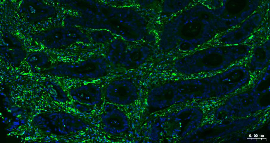
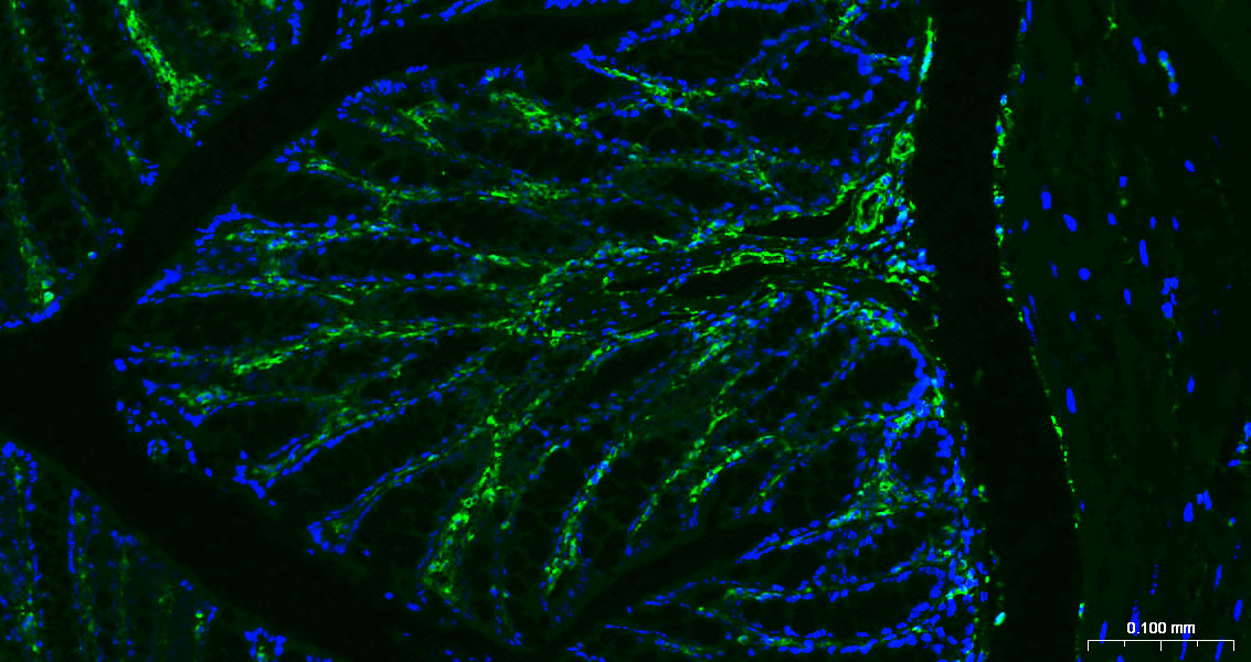
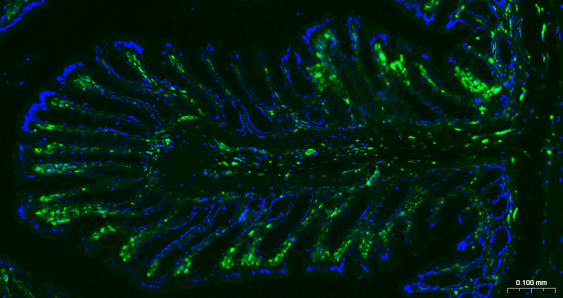
Paraformaldehyde-fixed, paraffin embedded Human Colon Cancer; Antigen retrieval by boiling in sodium citrate buffer (pH6.0) for 15 min; Antibody incubation with Vimentin Polyclonal Antibody, Unconjugated (bs-8533R) at 1:200 overnight at 4°C. Followed by conjugated Goat Anti-Rabbit IgG antibody (green, bs-0295G-BF488), DAPI (blue, C02-04002) was used to stain the cell nuclei.
Paraformaldehyde-fixed, paraffin embedded Mouse Colon; Antigen retrieval by boiling in sodium citrate buffer (pH6.0) for 15 min; Antibody incubation with Vimentin Polyclonal Antibody, Unconjugated (bs-8533R) at 1:200 overnight at 4°C. Followed by conjugated Goat Anti-Rabbit IgG antibody (green, bs-0295G-BF488), DAPI (blue, C02-04002) was used to stain the cell nuclei.
Paraformaldehyde-fixed, paraffin embedded Rat Colon; Antigen retrieval by boiling in sodium citrate buffer (pH6.0) for 15 min; Antibody incubation with Vimentin Polyclonal Antibody, Unconjugated (bs-8533R) at 1:200 overnight at 4°C. Followed by conjugated Goat Anti-Rabbit IgG antibody (green, bs-0295G-BF488), DAPI (blue, C02-04002) was used to stain the cell nuclei.
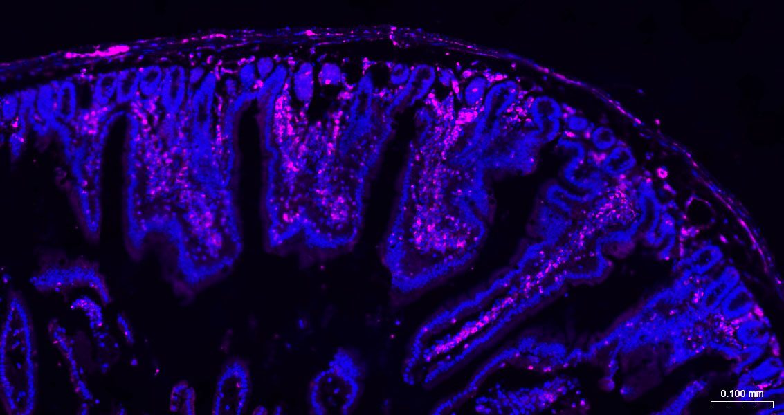
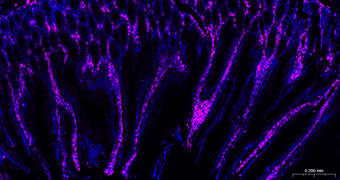
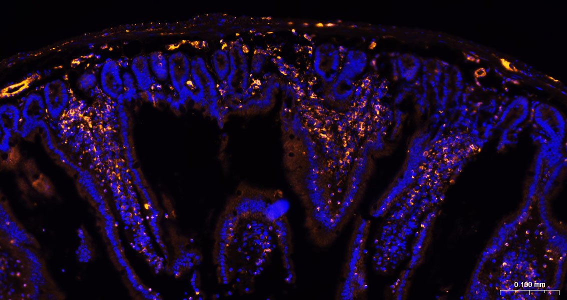
Paraformaldehyde-fixed, paraffin embedded Mouse Small Intestine; Antigen retrieval by boiling in sodium citrate buffer (pH6.0) for 15 min; The section was incubated with Vimentin Polyclonal Antibody, Unconjugated (bs-8533R) at 1:200 overnight at 4°C. Followed by conjugated Goat Anti-Rabbit IgG antibody (Rose Red, bs-40295G-BF647), DAPI (blue, C02-04002) was used to stain the cell nuclei.
Paraformaldehyde-fixed, paraffin embedded Rat Small Intestine; Antigen retrieval by boiling in sodium citrate buffer (pH6.0) for 15 min; The section was incubated with Vimentin Polyclonal Antibody, Unconjugated (bs-8533R) at 1:200 overnight at 4°C. Followed by conjugated Goat Anti-Rabbit IgG antibody (Rose Red, bs-40295G-BF647), DAPI (blue, C02-04002) was used to stain the cell nuclei.
Paraformaldehyde-fixed, paraffin embedded Human Kidney; Antigen retrieval by boiling in sodium citrate buffer (pH6.0) for 15 min; The section was incubated with Vimentin Polyclonal Antibody, Unconjugated (bs-8533R) at 1:200 overnight at 4°C. Followed by conjugated Goat Anti-Rabbit IgG antibody (Rose Red, bs-40295G-BF647), DAPI (blue, C02-04002) was used to stain the cell nuclei.
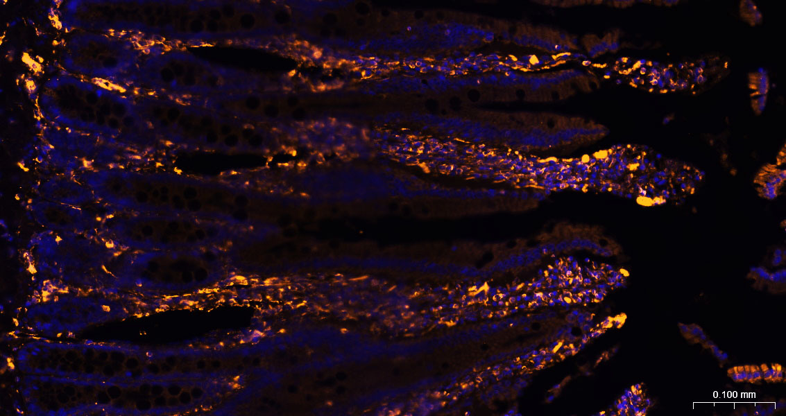
Paraformaldehyde-fixed, paraffin embedded Mouse Small Intestine ; Antigen retrieval by boiling in sodium citrate buffer (pH6.0) for 15min; The section was incubated with Vimentin Polyclonal Antibody, Unconjugated (bs-8533R) at 1:200 overnight at 4°C, followed by a conjugated Donkey Anti-Rabbit IgG antibody (bs-0295D-BF555) for 90 minutes, and DAPI for nuclei staining.
Paraformaldehyde-fixed, paraffin embedded Rat Small Intestine ; Antigen retrieval by boiling in sodium citrate buffer (pH6.0) for 15min; The section was incubated with Vimentin Polyclonal Antibody, Unconjugated (bs-8533R) at 1:200 overnight at 4°C, followed by a conjugated Donkey Anti-Rabbit IgG antibody (bs-0295D-BF555) for 90 minutes, and DAPI for nuclei staining.
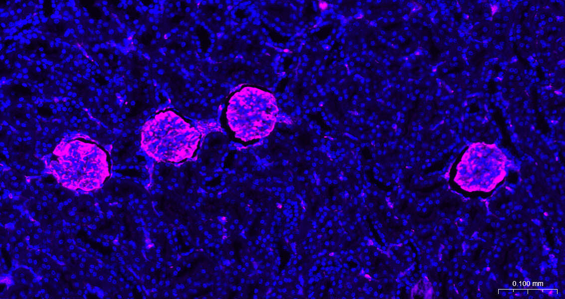
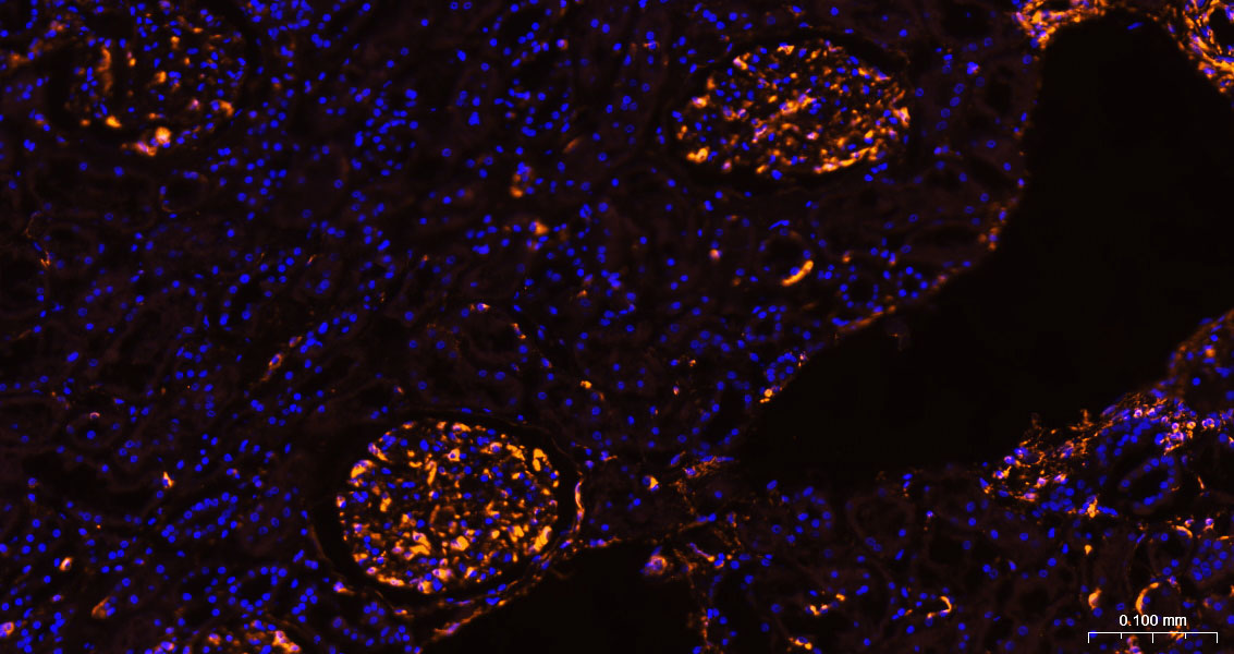
Paraformaldehyde-fixed, paraffin embedded Human Kidney ; Antigen retrieval by boiling in sodium citrate buffer (pH6.0) for 15min; The section was incubated with Vimentin Polyclonal Antibody, Unconjugated (bs-8533R) at 1:200 overnight at 4°C, followed by a conjugated Donkey Anti-Rabbit IgG antibody (bs-0295D-BF555) for 90 minutes, and DAPI for nuclei staining.
Paraformaldehyde-fixed, paraffin embedded Mouse Kidney ; Antigen retrieval by boiling in sodium citrate buffer (pH6.0) for 15min; The section was incubated with Vimentin Polyclonal Antibody, Unconjugated (bs-8533R) at 1:200 overnight at 4°C, followed by a conjugated Donkey Anti-Rabbit IgG antibody (bs-0295D-BF555) for 90 minutes, and DAPI for nuclei staining.
Paraformaldehyde-fixed, paraffin embedded Rat Kidney ; Antigen retrieval by boiling in sodium citrate buffer (pH6.0) for 15min; The section was incubated with Vimentin Polyclonal Antibody, Unconjugated (bs-8533R) at 1:200 overnight at 4°C, followed by a conjugated Donkey Anti-Rabbit IgG antibody (bs-0295D-BF555) for 90 minutes, and DAPI for nuclei staining.
Blank control:A549.
Primary Antibody (green line): Rabbit Anti-Vimentin antibody (bs-8533R)
Dilution: 1μg /10^6 cells;
Isotype Control Antibody (orange line): Rabbit IgG .
Secondary Antibody : Goat anti-rabbit IgG-AF488
Dilution: 1μg /test.
Protocol
The cells were fixed with 4% PFA (10min at room temperature)and then permeabilized with 90% ice-cold methanol for 20 min at -20℃. The cells were then incubated in 5%BSA to block non-specific protein-protein interactions for 30 min at room temperature .Cells stained with Primary Antibody for 30 min at room temperature. The secondary antibody used for 40 min at room temperature. Acquisition of 20,000 events was performed.