sales@bioss.com.cn
techsupport@bioss.com.cn
400-901-9800
Host: Rabbit
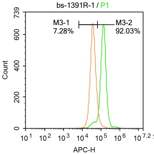
Target Protein: CHEK2 Rabbit pAb
IR: Immunogen Range:101-250/586
Clonality: Polyclonal
Isotype: IgG
Entrez Gene: 11200
Swiss Prot: O96017
Source: KLH conjugated synthetic peptide derived from human CHEK2:101-250/586
Purification: affinity purified by Protein A
Storage: 0.01M TBS (pH7.4) with 1% BSA, 0.02% Proclin300 and 50% Glycerol. Shipped at 4℃. Store at -20℃ for one year. Avoid repeated freeze/thaw cycles.
Background: In response to DNA damage and replication blocks, cell cycle progression is halted through the control of critical cell cycle regulators. The protein encoded by this gene is a cell cycle checkpoint regulator and putative tumor suppressor. It contains a forkhead-associated protein interaction domain essential for activation in response to DNA damage and is rapidly phosphorylated in response to replication blocks and DNA damage. When activated, the encoded protein is known to inhibit CDC25C phosphatase, preventing entry into mitosis, and has been shown to stabilize the tumor suppressor protein p53, leading to cell cycle arrest in G1. In addition, this protein interacts with and phosphorylates BRCA1, allowing BRCA1 to restore survival after DNA damage. Mutations in this gene have been linked with Li-Fraumeni syndrome, a highly penetrant familial cancer phenotype usually associated with inherited mutations in TP53. Also, mutations in this gene are thought to confer a predisposition to sarcomas, breast cancer, and brain tumors. This nuclear protein is a member of the CDS1 subfamily of serine/threonine protein kinases. Several transcript variants encoding different isoforms have been found for this gene. [provided by RefSeq, Apr 2012]
Size: 100ul
Concentration: 1mg/ml
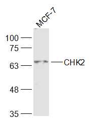
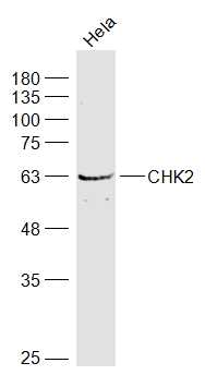
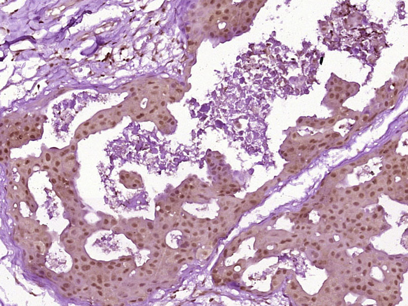
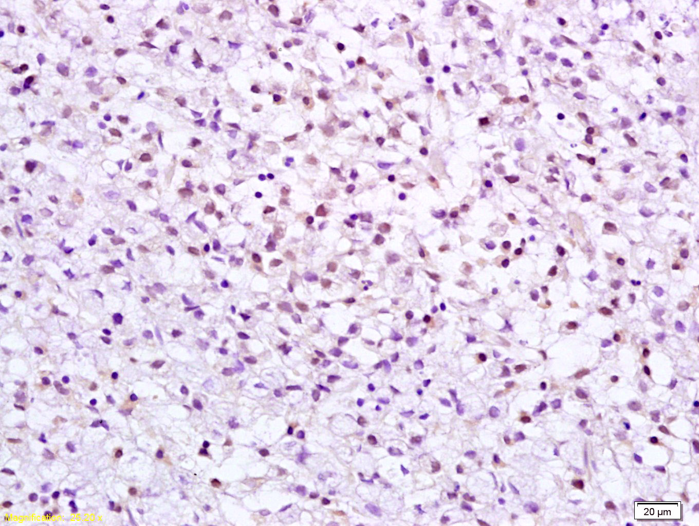
Applications: WB=1:500-2000,IHC-P=1:100-500,IHC-F=1:100-500,IF=1:100-500,Flow-Cyt=1ug/Tes
Cross Reactive Species: Human (predicted: Mouse,Rat,Rabbit,Cow,Dog,Horse)
For research use only. Not intended for diagnostic or therapeutic use.